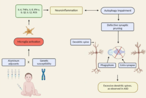
Microglial activation and increased microglial density observed in the dorsolateral prefrontal cortex in autism
Biol Psychiatry. 2010 Aug 15;68(4):368-76. doi: 10.1016/j.biopsych.2010.05.024.
Morgan JT1, Chana G, Pardo CA, Achim C, Semendeferi K, Buckwalter J, Courchesne E, Everall IP. Department of Neuroscience, School of Medicine, University of California, San Diego
BACKGROUND: In the neurodevelopmental disorder autism, several neuroimmune abnormalities have been reported. However, it is unknown whether microglial somal volume or density are altered in the cortex and whether any alteration is associated with age or other potential covariates.
METHODS: Microglia in sections from the dorsolateral prefrontal cortex of nonmacrencephalic male cases with autism (n = 13) and control cases (n = 9) were visualized via ionized calcium binding adapter molecule 1 immunohistochemistry. In addition to a neuropathological assessment, microglial cell density was stereologically estimated via optical fractionator and average somal volume was quantified via isotropic nucleator.
RESULTS: Microglia appeared markedly activated in 5 of 13 cases with autism, including 2 of 3 under age 6, and marginally activated in an additional 4 of 13 cases. Morphological alterations included somal enlargement, process retraction and thickening, and extension of filopodia from processes. Average microglial somal volume was significantly increased in white matter (p = .013), with a trend in gray matter (p = .098). Microglial cell density was increased in gray matter (p = .002). Seizure history did not influence any activation measure.
CONCLUSIONS: The activation profile described represents a neuropathological alteration in a sizeable fraction of cases with autism. Given its early presence, microglial activation may play a central role in the pathogenesis of autism in a substantial proportion of patients. Alternatively, activation may represent a response of the innate neuroimmune system to synaptic, neuronal, or neuronal network disturbances, or reflect genetic and/or environmental abnormalities impacting multiple cellular populations.
