GI

(digestive system) Group of organs stretching from the mouth to the anus, serving to breakdown foods, assimilate nutrients, and eliminate waste; in humans, the digestive system includes the gastrointestinal tract (mouth, esophagus, stomach, intestines, anus) and the accessory glands (liver, biliary tract, pancreas), and associated hormones and secretions. – National Institutes of Health

Excerpts:

“We found a functional architecture along the GBA that correlates with heterogeneity of ASD phenotypes, and it is characterized by ASD-associated amino acid, carbohydrate and lipid profiles predominantly encoded by microbial species in the genera Prevotella, Bifidobacterium, Desulfovibrio and Bacteroides and correlates with brain gene expression changes, restrictive dietary patterns and pro-inflammatory cytokine profiles.”

“We also show a strong association between temporal changes in microbiome composition and ASD phenotypes. In summary, we propose a framework to leverage multi-omic datasets from well-defined cohorts and investigate how the GBA influences ASD.”

More
  • June 26, 2023

Excerpts:

“Children with autism spectrum disorders often display dysregulated immune responses and related gastrointestinal symptoms.”

“Our study provides mechanistic insights into why children exposed to heightened inflammation in the womb might have an increased risk of developing inflammatory diseases in addition to neurodevelopmental disorders.”

More
  • December 7, 2021

Excerpt:
“Accumulating evidence implies the gut-brain axis as a pathway for MeHg harmful neurotoxic effects and a potential factor for later neurodegenerative disorders. The MeHg may induce a hormesis-related neuronal toxicity. Hormesis is an important redox dependent aging-associated neurodegenerative/ neuroprotective issue (Calabrese et al., 2010). The use of antioxidants, such as plant polyphenols (Calabrese et al., 2010; Leri et al., 2020) and protective nutrients (Oria et al., 2020) may be beneficial in reducing the MeHg-driven neuroinflammatory state and associated cell death with the interplay of the intestinal microbiota.”

More
  • November 3, 2020

“Background: Encephalitis, the inflammation of the brain, may be caused by an infection or an autoimmune reaction.”

“Conclusion: Gut microbiota disruption was observed in encephalitis patients, which manifested as pathogen dominance and health-promoting commensal depletion. Disease severity and brain damage may have associations with the gut microbiota or its metabolites.”

More
  • August 20, 2020

Excerpts:

“Herein, we will discuss the accumulating literature for ASD, giving special attention to the relevant aspects of factors that may be related to the neuroimmune interface in the development of ASD, including changes in neuroplasticity.”

Commentary on the article:

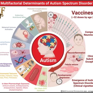
“The authors rightly highlight the newest challenging frontier of autism research: the neuroimmune axis alterations. These alterations are first evident in the cells early responsible for immune responses, as they are the precursors for macrophages, dendritic, and microglial cells: monocytes or peripheral blood mononuclear cells (PBMCs). These cells show strong dysfunctions in ASD children and are committed to a pro-inflammatory state, which in turn result in long-term immune alterations (4). In ASDs, altered PBMCs are responsible for elevated pro-inflammatory cytokine production. The up-regulation of inflammatory cytokines is also reflected in brain centers of autistic patients (5): the consequences are the induction of blood–brain barrier (the immunological interface between peripheral immune system and central nervous system) disruption. Changes in BBB permeability directly influence neural plasticity, connectivity and function, triggering impairments in social interaction, communication, and behavior (3). Immunological abnormalities also influence the gastrointestinal system and the microglial innate immune cells of the central nervous system (6). The authors also discuss the role of autoimmunity in the pathogenesis of autism. Familial or virus/bacteria-infected autoimmunity could be a risk factor for autism. Even if the exact cellular and molecular pathways responsible for the induction of neuroimmune alterations are still to be further clarify, a complex interaction among epigenetic and environmental risk factors (7) could trigger the neuroimmune abnormalities, such as abnormal neuron and glia responses.”

More
  • September 9, 2018

Excerpt:
“In this review article, we examine the connections between early disruption of the developing microbiome and gastrointestinal tract function, with particular regard to susceptibility to autism. The biological mechanisms that accompany individuals with autism are reviewed in this manuscript including immune system dysregulation, inflammation, oxidative stress, metabolic and methylation abnormalities as well as gastrointestinal distress.”

More
  • August 15, 2018

Excerpts:

“The likelihood of the child having ASD more than doubled among children with food allergy compared with those without food allergy; children with respiratory and skin allergy were also significantly more likely to have ASD, but at a lesser magnitude. While no sex difference was found for food allergy, boys with ASD were significantly more likely than girls with ASD to have respiratory and skin allergy.”

” It may be that GI dysfunction, seizures, and sleep disorder, in addition to food, respiratory, and skin allergies, are medical comorbidities that characterize the immune-mediated subtype of ASD.”

“In the Discussion section of their article, Xu and colleagues review other aspects of immune dysfunction reported in ASD, including abnormalities in peripheral immunoglobulins, imbalance of T-cell subsets, and increased levels of proinflammatory cytokines in postmortem brains of patients with ASD. Considering the significant association between food, respiratory, and skin allergy in children with ASD reported by Xu and colleagues, in conjunction with numerous studies documenting aspects of immune dysfunction in patients with ASD and specific animal models of ASD, evidence continues to mount that an immune-mediated subtype of ASD should continue to be pursued and defined.”

More
  • June 8, 2018

CONCLUSIONS AND RELEVANCE In a nationally representative sample of US children, a significant and positive association of common allergic conditions, in particular food allergy, with ASD was found. Further investigation is warranted to elucidate the causality and underlying mechanism

More
  • June 8, 2018

Abstract

There are similarities between the immune response following immunization with aluminum adjuvants and the immune response elicited by some helminthic parasites, including stimulation of immunoglobulin E (IgE) and eosinophilia. Immunization with aluminum adjuvants, as with helminth infection, induces a Th2 type cell mediated immune response, including eosinophilia, but does not induce an environment conducive to the induction of regulatory mechanisms. Helminths play a role in what is known as the hygiene hypothesis, which proposes that decreased exposure to microbes during a critical time in early life has resulted in the increased prevalence and morbidity of asthma and atopic disorders over the past few decades, especially in Western countries. In addition, gut and lung microbiome composition and their interaction with the immune system plays an important role in a properly regulated immune system. Disturbances in microbiome composition are a risk factor for asthma and allergies. We propose that immunization with aluminum adjuvants in general is not favorable for induction of regulatory mechanisms and, in the context of the hygiene hypothesis and microbiome theory, can be viewed as an amplifying factor and significant contributing risk factor for allergic diseases, especially in a genetically susceptible subpopulation.

More
  • May 3, 2018

Excerpt:
“The literature strongly supports that autism is most accurately seen as an acquired cellular detoxification deficiency syndrome with heterogeneous genetic predisposition that manifests pathophysiologic consequences of accumulated, run-away cellular toxicity. At a more general level, it is a form of a toxicant-induced loss of tolerance of toxins, and of chronic and sustained ER overload (“ER hyperstress”), contributing to neuronal and glial apoptosis via the unfolded protein response (UPR). Inherited risk of impaired cellular detoxification and circulating metal re-toxification in neurons and glial cells accompanied by chronic UPR is key.”

More
  • March 16, 2018