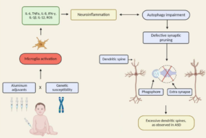
Discussion: The observation of predominantly intracellular aluminium in these tissues was novel and something similar has only previously been observed in cases of autism. The results suggest a strong inflammatory component in this case and support a role for aluminium in this rare and unusual case of CAA.
Astrocytes
A class of large neuroglial (macroglial) cells in the central nervous system – the largest and most numerous neuroglial cells in the brain and spinal cord. Astrocytes (from “star” cells) are irregularly shaped with many long processes, including those with “end feet” which form the glial (limiting) membrane and directly and indirectly contribute to the BLOOD-BRAIN BARRIER. They regulate the extracellular ionic and chemical environment, and “reactive astrocytes” (along with MICROGLIA) respond to injury. – NLM Medical Subject Headings, U.S. National Library of Medicine
Excerpt:
“[S]tudies linking IL-6 and autism, suggesting potential novel therapeutic pathways for a subtype of ASD individuals. This is the first report demonstrating that glial dysfunctions could contribute to non-syndromic autism pathophysiology using iPSCs modeling disease technology.”
Excerpt:
“Environmental mercury is neurotoxic at doses well below the current reference levels considered to be safe, with evidence of neurotoxicity in children exposed to environmental sources including fish consumption and ethylmercury-containing vaccines. Possible neurotoxic mechanisms of mercury include direct effects on sulfhydryl groups, pericytes and cerebral endothelial cells, accumulation within astrocytes, microglial activation, induction of chronic oxidative stress, activation of immune-inflammatory pathways and impairment of mitochondrial functioning. (Epi-)genetic factors which may increase susceptibility to the toxic effects of mercury in ASD include the following: a greater propensity of males to the long-term neurotoxic effects of postnatal exposure and genetic polymorphisms in glutathione transferases and other glutathione-related genes and in selenoproteins. Furthermore, immune and inflammatory responses to immunisations with mercury-containing adjuvants are strongly influenced by polymorphisms in the human leukocyte antigen (HLA) region and by genes encoding effector proteins such as cytokines and pattern recognition receptors. Some epidemiological studies investigating a possible relationship between high environmental exposure to methylmercury and impaired neurodevelopment have reported a positive dose-dependent effect.”
Excerpt: “Taken together, the results suggest a close link between oxidative stress neuroinflamation and degeneration in aluminium-fluoride toxicity.”
Abstract
Thimerosal generates ethylmercury in aqueous solution and is widely used as preservative. We have investigated the toxicology of Thimerosal in normal human astrocytes, paying particular attention to mitochondrial function and the generation of specific oxidants. We find that ethylmercury not only inhibits mitochondrial respiration leading to a drop in the steady state membrane potential, but also concurrent with these phenomena increases the formation of superoxide, hydrogen peroxide, and Fenton/Haber-Weiss generated hydroxyl radical. These oxidants increase the levels of cellular aldehyde/ketones. Additionally, we find a five-fold increase in the levels of oxidant damaged mitochondrial DNA bases and increases in the levels of mtDNA nicks and blunt-ended breaks. Highly damaged mitochondria are characterized by having very low membrane potentials, increased superoxide/hydrogen peroxide production, and extensively damaged mtDNA and proteins. These mitochondria appear to have undergone a permeability transition, an observation supported by the five-fold increase in Caspase-3 activity observed after Thimerosal treatment.
Excerpt:
“In summary, NF-κB is aberrantly expressed in orbitofrontal cortex in patients with ASC, as part of a putative molecular cascade leading to inflammation, especially of resident immune cells in brain regions associated with the behavioral and clinical symptoms of ASC.”
Excerpt:
“Significant cognitive deficits in water-maze learning were observed in the combined aluminum and squalene group (4.3 errors per trial) compared with the controls (0.2 errors per trial) after 20 wk. Apoptotic neurons were identified in aluminum-injected animals that showed significantly increased activated caspase-3 labeling in lumbar spinal cord (255%) and primary motor cortex (192%) compared with the controls. Aluminum-treated groups also showed significant motor neuron loss (35%) and increased numbers of astrocytes (350%) in the lumbar spinal cord. The findings suggest a possible role for the aluminum adjuvant in some neurological features associated with GWI and possibly an additional role for the combination of adjuvants.”
Exceprt:
” These results suggest that the IHg may be responsible for the increase in reactive glia.”
