Abstract
Autism spectrum disorder is a neurodevelopmental disorder of unknown aetiology. It is suggested to involve both genetic susceptibility and environmental factors including in the latter environmental toxins. Human exposure to the environmental toxin aluminium has been linked, if tentatively, to autism spectrum disorder. Herein we have used transversely heated graphite furnace atomic absorption spectrometry to measure, for the first time, the aluminium content of brain tissue from donors with a diagnosis of autism. We have also used an aluminium-selective fluor to identify aluminium in brain tissue using fluorescence microscopy. The aluminium content of brain tissue in autism was consistently high. The mean (standard deviation) aluminium content across all 5 individuals for each lobe were 3.82(5.42), 2.30(2.00), 2.79(4.05) and 3.82(5.17) μg/g dry wt. for the occipital, frontal, temporal and parietal lobes respectively. These are some of the highest values for aluminium in human brain tissue yet recorded and one has to question why, for example, the aluminium content of the occipital lobe of a 15 year old boy would be 8.74 (11.59) μg/g dry wt.? Aluminium-selective fluorescence microscopy was used to identify aluminium in brain tissue in 10 donors. While aluminium was imaged associated with neurones it appeared to be present intracellularly in microglia-like cells and other inflammatory non-neuronal cells in the meninges, vasculature, grey and white matter. The pre-eminence of intracellular aluminium associated with non-neuronal cells was a standout observation in autism brain tissue and may offer clues as to both the origin of the brain aluminium as well as a putative role in autism spectrum disorder.
Aluminum
Excerpt:
“The literature strongly supports that autism is most accurately seen as an acquired cellular detoxification deficiency syndrome with heterogeneous genetic predisposition that manifests pathophysiologic consequences of accumulated, run-away cellular toxicity. At a more general level, it is a form of a toxicant-induced loss of tolerance of toxins, and of chronic and sustained ER overload (“ER hyperstress”), contributing to neuronal and glial apoptosis via the unfolded protein response (UPR). Inherited risk of impaired cellular detoxification and circulating metal re-toxification in neurons and glial cells accompanied by chronic UPR is key.”
Abstract
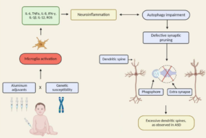
The conceptualisation of autistic spectrum disorder and Alzheimer’s disease has undergone something of a paradigm shift in recent years and rather than being viewed as single illnesses with a unitary pathogenesis and pathophysiology they are increasingly considered to be heterogeneous syndromes with a complex multifactorial aetiopathogenesis, involving a highly complex and diverse combination of genetic, epigenetic and environmental factors. One such environmental factor implicated as a potential cause in both syndromes is aluminium, as an element or as part of a salt, received, for example, in oral form or as an adjuvant. Such administration has the potential to induce pathology via several routes such as provoking dysfunction and/or activation of glial cells which play an indispensable role in the regulation of central nervous system homeostasis and neurodevelopment. Other routes include the generation of oxidative stress, depletion of reduced glutathione, direct and indirect reductions in mitochondrial performance and integrity, and increasing the production of proinflammatory cytokines in both the brain and peripherally. The mechanisms whereby environmental aluminium could contribute to the development of the highly specific pattern of neuropathology seen in Alzheimer’s disease are described. Also detailed are several mechanisms whereby significant quantities of aluminium introduced via immunisation could produce chronic neuropathology in genetically susceptible children. Accordingly, it is recommended that the use of aluminium salts in immunisations should be discontinued and that adults should take steps to minimise their exposure to environmental aluminium.
Excerpt:
“The most deficient element was zinc (92% in target and 20% in control), then – manganese (55% and 8%) and selenium (38% and 4%). In case of cooper study revealed excess concentration of this element only in target group in 50% of cases. The contaminations to heavy metals were detected in case of lead (78% and 16), mercury (43% and 10%) and cadmium (38% and 8%). The study statistical results indicated, that deficient concentrations of trace elements such as zinc, manganese, molybdenum and selenium in hair significantly linked with ASD (Kramer’s V was 0,740; 0,537; 0,333; 0,417 accordingly). In case of cooper we got excess levels of this element and this data was highly linked with autism spectrum disorder. We got high associations and significant values between of lead, mercury and cadmium concentrations and ASD. Study results indicate that there are significant differences of hair essential trace elements concentrations in children with autism spectrum disorder comparing with healthy children group. The result obtained also showed high contamination to heavy metals such as lead, mercury and cadmium in ASD children compared to healthy ones. So, our study demonstrated alteration in levels of toxic heavy metals and essential trace elements in children with autistic spectrum disorders as compared to healthy children. This suggests a possible pathophysiological role of heavy metals and trace elements in the genesis of symptoms of autism spectrum disorders.”
Excerpt: “Taken together, the results suggest a close link between oxidative stress neuroinflamation and degeneration in aluminium-fluoride toxicity.”
Abstract
The autism spectrum disorders (ASD) are a group of related neurodevelopmental disorders that have been increasing in incidence since the 1980s. Despite a considerable amount of data being collected from cases, a central mechanism has not been offered. A careful review of ASD cases discloses a number of events that adhere to an immunoexcitotoxic mechanism. This mechanism explains the link between excessive vaccination, use of aluminum and ethylmercury as vaccine adjuvants, food allergies, gut dysbiosis, and abnormal formation of the developing brain. It has now been shown that chronic microglial activation is present in autistic brains from age 5 years to age 44 years. A considerable amount of evidence, both experimental and clinical, indicates that repeated microglial activation can initiate priming of the microglia and that subsequent stimulation can produce an exaggerated microglial response that can be prolonged. It is also known that one phenotypic form of microglia activation can result in an outpouring of neurotoxic levels of the excitotoxins, glutamate and quinolinic acid. Studies have shown that careful control of brain glutamate levels is essential to brain pathway development and that excesses can result in arrest of neural migration, as well as dendritic and synaptic loss. It has also been shown that certain cytokines, such as TNF-alpha, can, via its receptor, interact with glutamate receptors to enhance the neurotoxic reaction. To describe this interaction I have coined the term immunoexcitotoxicity, which is described in this article.
Excerpts:
“Animal models of neurological disease plainly suggest that the ubiquitous presence of Al in human beings implicates Al toxicants as causally involved in Lou Gehrig’s disease (ALS), Alzheimer’s disease and autism spectrum disorders.”
“All these findings plausibly implicate Al adjuvants in pediatric vaccines as causal factors contributing to increased rates of autism spectrum disorders in countries where multiple doses are almost universally administered.“
Results
The CDDS and IDEA data sets are qualitatively consistent in suggesting a strong increase in autism prevalence over recent decades. The quantitative comparison of IDEA snapshot and constant-age tracking trend slopes suggests that ~75-80% of the tracked increase in autism since 1988 is due to an actual increase in the disorder rather than to changing diagnostic criteria. Most of the suspected environmental toxins examined have flat or decreasing temporal trends that correlate poorly to the rise in autism. Some, including lead, organochlorine pesticides and vehicular emissions, have strongly decreasing trends. Among the suspected toxins surveyed, polybrominated diphenyl ethers, aluminum adjuvants, and the herbicide glyphosate have increasing trends that correlate positively to the rise in autism.
Abstract
Autism spectrum disorders (ASDs) are complex, heterogeneous disorders caused by an interaction between genetic vulnerability and environmental factors. In an effort to better target the underlying roots of ASD for diagnosis and treatment, efforts to identify reliable biomarkers in genetics, neuroimaging, gene expression, and measures of the body’s metabolism are growing. For this article, we review the published studies of potential biomarkers in autism and conclude that while there is increasing promise of finding biomarkers that can help us target treatment, there are none with enough evidence to support routine clinical use unless medical illness is suspected. Promising biomarkers include those for mitochondrial function, oxidative stress, and immune function. Genetic clusters are also suggesting the potential for useful biomarkers.
Excerpt: “A recent review assessed the research on physiological abnormalities associated with ASD (44). The authors identified four main mechanisms that have been increasingly studied during the past decade: immunologic/inflammation, oxidative stress, environmental toxicants, and mitochondrial abnormalities. In addition, there is accumulating research on the lipid, GI systems, microglial activation, and the microbiome, and how these can also contribute to generating biomarkers associated with ASD (45, 46).
Pathways are interconnected with a defect in one likely leading to dysfunction in others. Many metabolic disorders can lead to endpoints such as impaired methylation, sulfuration, and detoxification pathways and nutritional deficiencies. Mitochondrial dysfunction, environmental risk factors, metabolic imbalances, and genetic susceptibility can all lead to oxidative stress (47), which in turn leads to inflammation, damaged cell membranes, autoimmunity (48), impaired methylation (49), cell death (48), and neurological deficits (50). The brain is highly vulnerable to oxidative stress (51), particularly in children (52) during the early part of development (47). As environmental events and metabolic imbalances affect oxidative stress and methylation, they also can affect the expression of genes.”
Abstract
Regulatory T cells play a critical role in the immune response to vaccination, but there is only a limited understanding of the response of regulatory T cells to aluminum adjuvants and the vaccines that contain them. Available studies in animal models show that although induced T regulatory cells may be induced concomitantly with effector T cells following aluminum-adjuvanted vaccination, they are unable to protect against sensitization, suggesting that under the Th2 immune-stimulating effects of aluminum adjuvants, Treg cells may be functionally compromised. Allergic diseases are characterized by immune dysregulation, with increases in IL-4 and IL-6, both of which exert negative effects on Treg function. For individuals with a genetic predisposition, the beneficial influence of adjuvants on immune responsiveness may be accompanied by immune dysregulation, leading to allergic diseases. This review examines aspects of the regulatory T cell response to aluminum-adjuvanted immunization and possible genetic susceptibility factors related to that response.
