Excerpt:
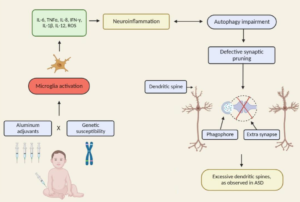
“Environmental mercury is neurotoxic at doses well below the current reference levels considered to be safe, with evidence of neurotoxicity in children exposed to environmental sources including fish consumption and ethylmercury-containing vaccines. Possible neurotoxic mechanisms of mercury include direct effects on sulfhydryl groups, pericytes and cerebral endothelial cells, accumulation within astrocytes, microglial activation, induction of chronic oxidative stress, activation of immune-inflammatory pathways and impairment of mitochondrial functioning. (Epi-)genetic factors which may increase susceptibility to the toxic effects of mercury in ASD include the following: a greater propensity of males to the long-term neurotoxic effects of postnatal exposure and genetic polymorphisms in glutathione transferases and other glutathione-related genes and in selenoproteins. Furthermore, immune and inflammatory responses to immunisations with mercury-containing adjuvants are strongly influenced by polymorphisms in the human leukocyte antigen (HLA) region and by genes encoding effector proteins such as cytokines and pattern recognition receptors. Some epidemiological studies investigating a possible relationship between high environmental exposure to methylmercury and impaired neurodevelopment have reported a positive dose-dependent effect.”
Endothelial Cells
Highly specialized epithelial cells that line the heart; blood vellels; and lymph vessels, forming the endothelium. They are polygonal in shape and joined together by tight jumctions. The tight junctions allow for variable permeability to specific macromolecules that are transported across the endothelial layer. – NLM Medical Subject Headings
Conclusions: HgCl2 stimulates VEGF and IL-6 release from human mast cells. This phenomenon could disrupt the blood-brain-barrier and permit brain inflammation. As a result, the findings of the present study provide a biological mechanism for how low levels of mercury may contribute to ASD pathogenesis.
