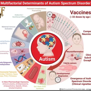
Conclusion: The totality of evidence supports a multifactorial model of ASD in which genetic predisposition, neuroimmune biology, environmental toxicants, perinatal stressors, and iatrogenic exposures converge to produce the phenotype of a post-encephalitic state. Combination and early-timed routine childhood vaccination constitutes the most significant modifiable risk factor for ASD, supported by convergent mechanistic, clinical, and epidemiologic findings, and characterized by intensified use, the clustering of multiple doses during critical neurodevelopmental windows, and the lack of research on the cumulative safety of the full pediatric schedule. As ASD prevalence continues to rise at an unprecedented pace, clarifying the risks associated with cumulative vaccine dosing and timing remains an urgent public health priority.
Neurons
Basic cellular units of nervous tissue; each neuron consists of a body, an axon, and dendrites and their purpose is to receive, conduct, and transmit impulses in the nervous system. – CRISP Thesaurus, National Institutes of Health
Highlights
•Exposure to ethylmercury (EtHG) poses neurotoxic risks in children.
•Behavioral abnormalities were induced by EtHg exposure in neonatal mice.
•Changes in neurite length and BDNF expression were observed.
•EtHg induces physiological changes in the brain by activating microglia.
Excerpt
“Our findings revealed that EtHg exposure led to significant alterations in brain development, including increased brain size and cortical thickness. These structural changes were accompanied by notable impairments in social interactions and behavioral patterns. Further analysis indicated that these effects were likely mediated by increased microglial activation and elevated BDNF expression in the cerebral cortex. Overall, our study suggests that EtHg disrupts neurodevelopment by activating microglia, leading to physiological and morphological changes in the brain.
Excerpt:
“Accumulating evidence implies the gut-brain axis as a pathway for MeHg harmful neurotoxic effects and a potential factor for later neurodegenerative disorders. The MeHg may induce a hormesis-related neuronal toxicity. Hormesis is an important redox dependent aging-associated neurodegenerative/ neuroprotective issue (Calabrese et al., 2010). The use of antioxidants, such as plant polyphenols (Calabrese et al., 2010; Leri et al., 2020) and protective nutrients (Oria et al., 2020) may be beneficial in reducing the MeHg-driven neuroinflammatory state and associated cell death with the interplay of the intestinal microbiota.”
Excerpts:
“Aberrant migration of inhibitory interneurons can alter the formation of cortical circuitry and lead to severe neurologic disorders including epilepsy, autism, and schizophrenia.”
“These findings highlight key roles for JNK signaling in leading process branching, nucleokinesis, and the trafficking of centrosomes and cilia during interneuron migration, and further implicates JNK signaling as an important mediator of cortical development.”
Excerpt:
“The literature strongly supports that autism is most accurately seen as an acquired cellular detoxification deficiency syndrome with heterogeneous genetic predisposition that manifests pathophysiologic consequences of accumulated, run-away cellular toxicity. At a more general level, it is a form of a toxicant-induced loss of tolerance of toxins, and of chronic and sustained ER overload (“ER hyperstress”), contributing to neuronal and glial apoptosis via the unfolded protein response (UPR). Inherited risk of impaired cellular detoxification and circulating metal re-toxification in neurons and glial cells accompanied by chronic UPR is key.”
Excerpt:
“[S]tudies linking IL-6 and autism, suggesting potential novel therapeutic pathways for a subtype of ASD individuals. This is the first report demonstrating that glial dysfunctions could contribute to non-syndromic autism pathophysiology using iPSCs modeling disease technology.”
Excerpts:
“…several large scale epidemiological studies have recently linked prenatal air pollution exposure with an increased risk of neurodevelopmental disorders such as autism spectrum disorder (ASD).”
“We have demonstrated that prenatal exposure to DEP in mice, i.e., to the pregnant dams throughout gestation, results in a persistent vulnerability to behavioral deficits in adult offspring, especially in males, which is intriguing given the greater incidence of ASD in males to females (∼4:1).”
“DEP exposure increased inflammatory cytokine protein and altered the morphology of microglia, consistent with activation or a delay in maturation, only within the embryonic brains of male mice…”
“Consistent with this hypothesis, we found increased microglial-neuronal interactions in male offspring that received DEP compared to all other groups. Taken together, these data suggest a mechanism by which prenatal exposure to environmental toxins may affect microglial development and long-term function, and thereby contribute to the risk of neurodevelopmental disorders.”
Abstract
In this section, I explore the effects of mercury and inflammation on transsulfuration reactions, which can lead to elevations in androgens, and how this might relate to the male preponderance of autism spectrum disorders (ASD). It is known that mercury interferes with these biochemical reactions and that chronically elevated androgen levels also enhance the neurodevelopmental effects of excitotoxins. Both androgens and glutamate alter neuronal and glial calcium oscillations, which are known to regulate cell migration, maturation, and final brain cytoarchitectural structure. Studies have also shown high levels of DHEA and low levels of DHEA-S in ASD, which can result from both mercury toxicity and chronic inflammation. Chronic microglial activation appears to be a hallmark of ASD. Peripheral immune stimulation, mercury, and elevated levels of androgens can all stimulate microglial activation. Linked to both transsulfuration problems and chronic mercury toxicity are elevations in homocysteine levels in ASD patients. Homocysteine and especially its metabolic products are powerful excitotoxins. Intimately linked to elevations in DHEA, excitotoxicity and mercury toxicity are abnormalities in mitochondrial function. A number of studies have shown that reduced energy production by mitochondria greatly enhances excitotoxicity. Finally, I discuss the effects of chronic inflammation and elevated mercury levels on glutathione and metallothionein.
Excerpt:
“Our findings suggest that chemicals and genetic mutations that impair topoisomerases could commonly contribute to ASD and other neurodevelopmental disorders.”
Excerpt:
“In summary, NF-κB is aberrantly expressed in orbitofrontal cortex in patients with ASC, as part of a putative molecular cascade leading to inflammation, especially of resident immune cells in brain regions associated with the behavioral and clinical symptoms of ASC.”
