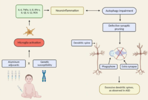
Excerpt:
“Our data thus demonstrate a negative neurodevelopmental impact of perinatal TM exposure which appears to be both strain- and sex-dependent.”
Oxidative Stress
Excerpt:
“In conclusion, these data suggest that thimerosal induced U937 activation via oxidative stress from mitochondrial stores and mitochondrial membrane depolarization with a primordial effect of thiol groups.”
Excerpt:
“Exposure to oxidative stress via the sulfhydryl reagent thimerosal resulted in a greater decrease in the GSH/GSSG ratio and increase in free radical generation in autism compared to control cells. Acute exposure to physiological levels of nitric oxide decreased mitochondrial membrane potential to a greater extent in the autism LCLs, although GSH/GSSG and ATP concentrations were similarly decreased in both cell lines. These results suggest that the autism LCLs exhibit a reduced glutathione reserve capacity in both cytosol and mitochondria that may compromise antioxidant defense and detoxification capacity under prooxidant conditions.”
Shows a potential link between mercury and the autopsied brains of young people with autism. A marker for oxidative stress was 68.9% higher in autistic brain issue than controls (a statistically significant result), while mercury levels were 68.2% higher.
Excerpt:
“The preliminary data suggest a need for more extensive studies of oxidative stress, its relationship to the environmental factors and its possible attenuation by antioxidants in autism.”
Excerpts:
“Such special cases suggest that the pathophysiology of autism may comprise pathways that are directly or indirectly involved in mitochondrial energy production…”
Excerpt:
“Exposure to environmental toxins is the likely etiology for MtD in autism. This dysfunction then contributes to a number of diagnostic symptoms and comorbidities observed in autism including: cognitive impairment, language deficits, abnormal energy metabolism, chronic gastrointestinal problems, abnormalities in fatty acid oxidation, and increased oxidative stress. MtD and oxidative stress may also explain the high male to female ratio found in autism due to increased male vulnerability to these dysfunctions.”
Excerpt:
“In autism, over-zealous neuroinflammatory responses could not only influence neural developmental processes, but may more significantly impair neural signaling involved in cognition in an ongoing fashion.”
Exceprts:
We also discuss evidence implicating oxidative stress, neuroglial activation and neuroimmunity in autism.
“Oxidative stress is another possible cause of Purkinje cell loss and other neuroanatomical changes described in autistic brains (reviewed in (37, 113)). Oxidative stress occurs when the levels of reactive oxygen species exceed the antioxidant capacities of a cell, often leading to cell death. Because of its very high oxygen demands and limited anti-oxidant capacity, the brain is thought to be relatively vulnerable to oxidative stress (111). Several studies have shown decreased levels of antioxidants such as superoxide dismutase, transferrin and ceruloplasmin in the blood or serum of patients with ASD (38, 108, 222). Significant elevations in biomarker profiles indicating increased oxidative stress, such as increased lipid peroxidation, have also been documented in autism (38, 107, 229).Interestingly, in one report the alterations in antioxidant proteins were linked specifically to regressive autism, suggesting a postnatal environmental effect (38). Polymorphisms in metabolic pathway genes may contribute to the increased oxidative stress in autism (108). Advanced glycationend products have also been reported to be elevated in both the brain tissue and serum of autistic patients, a change which can also lead to increased oxidative damage (23,110).”
Abstract
According to the Autism Society of America, autism is now considered to be an epidemic. The increase in the rate of autism revealed by epidemiological studies and government reports implicates the importance of external or environmental factors that may be changing. This article discusses the evidence for the case that some children with autism may become autistic from neuronal cell death or brain damage sometime after birth as result of insult; and addresses the hypotheses that toxicity and oxidative stress may be a cause of neuronal insult in autism. The article first describes the Purkinje cell loss found in autism, Purkinje cell physiology and vulnerability, and the evidence for postnatal cell loss. Second, the article describes the increased brain volume in autism and how it may be related to the Purkinje cell loss. Third, the evidence for toxicity and oxidative stress is covered and the possible involvement of glutathione is discussed. Finally, the article discusses what may be happening over the course of development and the multiple factors that may interplay and make these children more vulnerable to toxicity, oxidative stress, and neuronal insult.
Excerpt:
“The metabolic results indicated that plasma methionine and the ratio of S-adenosylmethionine (SAM) to S-adenosylhomocysteine (SAH), an indicator of methylation capacity, were significantly decreased in the autistic children relative to age-matched controls. In addition, plasma levels of cysteine, glutathione, and the ratio of reduced to oxidized glutathione, an indication of antioxidant capacity and redox homeostasis, were significantly decreased. Differences in allele frequency and/or significant gene-gene interactions were found for relevant genes encoding the reduced folate carrier (RFC 80G > A), transcobalamin II (TCN2 776G > C), catechol-O-methyltransferase (COMT 472G > A), methylenetetrahydrofolate reductase (MTHFR 677C > T and 1298A > C), and glutathione-S-transferase (GST M1). We propose that an increased vulnerability to oxidative stress (endogenous or environmental) may contribute to the development and clinical manifestations of autism.”
