Excerpt:
“The literature strongly supports that autism is most accurately seen as an acquired cellular detoxification deficiency syndrome with heterogeneous genetic predisposition that manifests pathophysiologic consequences of accumulated, run-away cellular toxicity. At a more general level, it is a form of a toxicant-induced loss of tolerance of toxins, and of chronic and sustained ER overload (“ER hyperstress”), contributing to neuronal and glial apoptosis via the unfolded protein response (UPR). Inherited risk of impaired cellular detoxification and circulating metal re-toxification in neurons and glial cells accompanied by chronic UPR is key.”
Review
Conclusions
Results of the current meta-analysis revealed that mercury is an important causal factor in the etiology of ASD. It seems that the detoxification and excretory mechanisms are impaired in ASD patients which lead to accumulation of mercury in the body. Future additional studies on mercury levels in different tissues of ASD patients should be undertaken.
Conclusion The literature reviewed here suggests a link between abnormalities in mitochondrial homeostasis and ASD and provides biochemical and genetic evidence to support a role for mitochondrial dysfunction in the pathogenesis of the autism phenotype.
Excerpts:
“The difference in manifested toxicity of MeHg and EtHg are likely the result of the differences in exposure, metabolism, and elimination from the body, rather than differences in mechanisms of action between the two.”
“Summary and Conclusions
There are many commonalities/similarities in the mechanisms of toxic action of methylmercury and ethylmercury (from thimerosal)… Evidence for the similarity of the various mechanisms of toxicity include the following:
• Both MeHg and EtHg bind to the amino acid cysteine (Clarkson 1995; Wu et al. 2008)…
• Both decrease glutathione activity, thus providing less protection from the oxidative stress caused by MeHg and EtHg (Carocci et al. 2014; Ndountse and Chan (2008); Choi et al. 1996; Franco et al. 2006; Mori et al. 2007; Muller et al. 2001; Ndountse and Chan 2008; Wu et al. 2008)…
• Both disrupt glutamate homeostasis (Farina et al. 2003a, b; Manfroi et al. 2004; Mutkus et al. 2005; Yin et al. 2007).
• Both cause oxidative stress/creation of ROS (Dreiem and Seegal 2007; Garg and Chang 2006; Myhre et al. 2003; Sharpe et al. 2012; Yin et al. 2007)…
• Both cause effects on receptor binding/neurotransmitter release involving one or more transmitters (Basu et al. 2008; Coccini et al. 2000; Cooper et al. 2003; Fonfria et al. 2001; Ida-Eto et al. 2011; Ndountse and Chan 2008; Yuan and Atchison 2003).
• Both cause DNA damage or impair DNA synthesis (Burke et al. 2006; Sharpe et al. 2012; Wu et al. 2008).”
Exerpt:
“Autism spectrum disorder (ASD) is over four times more prevalent in males compared to females.”
Abstract
In this section, I explore the effects of mercury and inflammation on transsulfuration reactions, which can lead to elevations in androgens, and how this might relate to the male preponderance of autism spectrum disorders (ASD). It is known that mercury interferes with these biochemical reactions and that chronically elevated androgen levels also enhance the neurodevelopmental effects of excitotoxins. Both androgens and glutamate alter neuronal and glial calcium oscillations, which are known to regulate cell migration, maturation, and final brain cytoarchitectural structure. Studies have also shown high levels of DHEA and low levels of DHEA-S in ASD, which can result from both mercury toxicity and chronic inflammation. Chronic microglial activation appears to be a hallmark of ASD. Peripheral immune stimulation, mercury, and elevated levels of androgens can all stimulate microglial activation. Linked to both transsulfuration problems and chronic mercury toxicity are elevations in homocysteine levels in ASD patients. Homocysteine and especially its metabolic products are powerful excitotoxins. Intimately linked to elevations in DHEA, excitotoxicity and mercury toxicity are abnormalities in mitochondrial function. A number of studies have shown that reduced energy production by mitochondria greatly enhances excitotoxicity. Finally, I discuss the effects of chronic inflammation and elevated mercury levels on glutathione and metallothionein.
Abstract
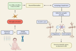
The autism spectrum disorders (ASD) are a group of related neurodevelopmental disorders that have been increasing in incidence since the 1980s. Despite a considerable amount of data being collected from cases, a central mechanism has not been offered. A careful review of ASD cases discloses a number of events that adhere to an immunoexcitotoxic mechanism. This mechanism explains the link between excessive vaccination, use of aluminum and ethylmercury as vaccine adjuvants, food allergies, gut dysbiosis, and abnormal formation of the developing brain. It has now been shown that chronic microglial activation is present in autistic brains from age 5 years to age 44 years. A considerable amount of evidence, both experimental and clinical, indicates that repeated microglial activation can initiate priming of the microglia and that subsequent stimulation can produce an exaggerated microglial response that can be prolonged. It is also known that one phenotypic form of microglia activation can result in an outpouring of neurotoxic levels of the excitotoxins, glutamate and quinolinic acid. Studies have shown that careful control of brain glutamate levels is essential to brain pathway development and that excesses can result in arrest of neural migration, as well as dendritic and synaptic loss. It has also been shown that certain cytokines, such as TNF-alpha, can, via its receptor, interact with glutamate receptors to enhance the neurotoxic reaction. To describe this interaction I have coined the term immunoexcitotoxicity, which is described in this article.
Conclusion
There is now sufficient evidence from both human and animal studies showing that cumulative exposure to aluminium adjuvants is not as benign as previously assumed. Given that vaccines are the only medical intervention that we attempt to deliver to every living human on earth and that by far the largest target population for vaccination are healthy children, a better appreciation and understanding of vaccine adjuvant risks appears warranted.
Excerpt:
“Evidence from such studies point to a half-life of inorganic mercury in human brains of several years to several decades.”
Excerpt:
“These findings suggest that infantile zinc- and magnesium-deficiency and/or toxic metal burdens may be critical and induce epigenetic alterations in the genes and genetic regulation mechanisms of neurodevelopment in the autistic children, and demonstrate that a time factor “infantile window” is also critical for neurodevelopment and probably for therapy. Thus, early metallomics analysis may lead to early screening/estimation and treatment/prevention for the autistic neurodevelopment disorders.”
