“Conclusion
We found that blood mercury levels at late pregnancy and early childhood were associated with more autistic behaviors in children at 5 years of age. Further study on the long-term effects of mercury exposure is recommended.”
Thimerosal
Abstract
Environmental factors have been implicated in the etiology of autism spectrum disorder (ASD); however, the role of heavy metals has not been fully defined. This study investigated whether blood levels of mercury, arsenic, cadmium, and lead of children with ASD significantly differ from those of age- and sex-matched controls. One hundred eighty unrelated children with ASD and 184 healthy controls were recruited. Data showed that the children with ASD had significantly (p < 0.001) higher levels of mercury and arsenic and a lower level of cadmium. The levels of lead did not differ significantly between the groups. The results of this study are consistent with numerous previous studies, supporting an important role for heavy metal exposure, particularly mercury, in the etiology of ASD. It is desirable to continue future research into the relationship between ASD and heavy metal exposure.
Abstract
Autism spectrum disorder (ASD) is a complex neurodevelopmental disorder that affects social, communication, and behavioral development. Recent evidence supported but also questioned the hypothetical role of compounds containing mercury (Hg) as contributors to the development of ASD. Specific alterations in the urinary excretion of porphyrin-containing ring catabolites have been associated with exposure to Hg in ASD patients. In the present study, the level of urinary porphyrins, as biomarkers of Hg toxicity in children with ASD, was evaluated, and its correlation with severity of the autistic behavior further explored. A total of 100 children was enrolled in the present study. They were classified into three groups: children with ASD (40), healthy controls (40), and healthy siblings of the ASD children (20). Children with ASD were diagnosed using DSM-IV-TR, ADI-R, and CARS tests. Urinary porphyrins were evaluated within the three groups using high-performance liquid chromatography (HPLC), after plasma evaluation of mercury (Hg) and lead (Pb) in the same groups. Results showed that children with ASD had significantly higher levels of Hg, Pb, and the porphyrins pentacarboxyporphyrin, coproporphyrin, precoproporphyrin, uroporphyrins, and hexacarboxyporphyrin compared to healthy controls and healthy siblings of the ASD children. However, there was no significant statistical difference in the level of heptacarboxyporphyrin among the three groups, while a significant positive correlation between the levels of coproporphyrin and precoproporphyrin and autism severity was observed. Mothers of ASD children showed a higher percentage of dental amalgam restorations compared to the mothers of healthy controls suggesting that high Hg levels in children with ASD may relate to the increased exposure to Hg from maternal dental amalgam during pregnancy and lactation. The results showed that the ASD children in the present study had increased blood Hg and Pb levels compared with healthy control children indicating that disordered porphyrin metabolism might interfere with the pathology associated with the autistic neurologic phenotype. The present study indicates that coproporphyrin and precoproporhyrin may be utilized as possible biomarkers for heavy metal exposure and autism severity in children with ASD.
Excerpt:
“We have reanalyzed the data set originally reported by Ip et al. in 2004 and have found that the original p value was in error and that a significant relation does exist between the blood levels of mercury and diagnosis of an autism spectrum disorder. Moreover, the hair sample analysis results offer some support for the idea that persons with autism may be less efficient and more variable at eliminating mercury from the blood.”
Excerpt:
“Levels of serum neurokinin A and BHg were measured in 84 children with ASD, aged between 3 and 10 years, and 84 healthy-matched children. There was a positive linear relationship between the Childhood Autism Rating Scale (CARS) and both serum neurokinin A and BHg. ASD children had significantly higher levels of serum neurokinin A than healthy controls (P < 0.001). Increased levels of serum neurokinin A and BHg were respectively found in 54.8 % and 42.9 % of the two groups. There was significant and positive linear relationship between levels of serum neurokinin A and BHg in children with moderate and severe ASD, but not in healthy control children. It was found that 78.3 % of the ASD patients with increased serum levels of neurokinin A had elevated BHg levels (P < 0.001). Neuroinflammation, with increased levels of neurokinin A, is seen in some children with ASD, and may be caused by elevated BHg levels. Further research is recommended to determine the pathogenic role of increased levels of serum neurokinin A and BHg in ASD. The therapeutic role of tachykinin receptor antagonists, a potential new class of anti-inflammatory medications, and Hg chelators, should also be studied in ASD."
Excerpts:
“The difference in manifested toxicity of MeHg and EtHg are likely the result of the differences in exposure, metabolism, and elimination from the body, rather than differences in mechanisms of action between the two.”
“Summary and Conclusions
There are many commonalities/similarities in the mechanisms of toxic action of methylmercury and ethylmercury (from thimerosal)… Evidence for the similarity of the various mechanisms of toxicity include the following:
• Both MeHg and EtHg bind to the amino acid cysteine (Clarkson 1995; Wu et al. 2008)…
• Both decrease glutathione activity, thus providing less protection from the oxidative stress caused by MeHg and EtHg (Carocci et al. 2014; Ndountse and Chan (2008); Choi et al. 1996; Franco et al. 2006; Mori et al. 2007; Muller et al. 2001; Ndountse and Chan 2008; Wu et al. 2008)…
• Both disrupt glutamate homeostasis (Farina et al. 2003a, b; Manfroi et al. 2004; Mutkus et al. 2005; Yin et al. 2007).
• Both cause oxidative stress/creation of ROS (Dreiem and Seegal 2007; Garg and Chang 2006; Myhre et al. 2003; Sharpe et al. 2012; Yin et al. 2007)…
• Both cause effects on receptor binding/neurotransmitter release involving one or more transmitters (Basu et al. 2008; Coccini et al. 2000; Cooper et al. 2003; Fonfria et al. 2001; Ida-Eto et al. 2011; Ndountse and Chan 2008; Yuan and Atchison 2003).
• Both cause DNA damage or impair DNA synthesis (Burke et al. 2006; Sharpe et al. 2012; Wu et al. 2008).”
Excerpt:
“The most deficient element was zinc (92% in target and 20% in control), then – manganese (55% and 8%) and selenium (38% and 4%). In case of cooper study revealed excess concentration of this element only in target group in 50% of cases. The contaminations to heavy metals were detected in case of lead (78% and 16), mercury (43% and 10%) and cadmium (38% and 8%). The study statistical results indicated, that deficient concentrations of trace elements such as zinc, manganese, molybdenum and selenium in hair significantly linked with ASD (Kramer’s V was 0,740; 0,537; 0,333; 0,417 accordingly). In case of cooper we got excess levels of this element and this data was highly linked with autism spectrum disorder. We got high associations and significant values between of lead, mercury and cadmium concentrations and ASD. Study results indicate that there are significant differences of hair essential trace elements concentrations in children with autism spectrum disorder comparing with healthy children group. The result obtained also showed high contamination to heavy metals such as lead, mercury and cadmium in ASD children compared to healthy ones. So, our study demonstrated alteration in levels of toxic heavy metals and essential trace elements in children with autistic spectrum disorders as compared to healthy children. This suggests a possible pathophysiological role of heavy metals and trace elements in the genesis of symptoms of autism spectrum disorders.”
Abstract
In this section, I explore the effects of mercury and inflammation on transsulfuration reactions, which can lead to elevations in androgens, and how this might relate to the male preponderance of autism spectrum disorders (ASD). It is known that mercury interferes with these biochemical reactions and that chronically elevated androgen levels also enhance the neurodevelopmental effects of excitotoxins. Both androgens and glutamate alter neuronal and glial calcium oscillations, which are known to regulate cell migration, maturation, and final brain cytoarchitectural structure. Studies have also shown high levels of DHEA and low levels of DHEA-S in ASD, which can result from both mercury toxicity and chronic inflammation. Chronic microglial activation appears to be a hallmark of ASD. Peripheral immune stimulation, mercury, and elevated levels of androgens can all stimulate microglial activation. Linked to both transsulfuration problems and chronic mercury toxicity are elevations in homocysteine levels in ASD patients. Homocysteine and especially its metabolic products are powerful excitotoxins. Intimately linked to elevations in DHEA, excitotoxicity and mercury toxicity are abnormalities in mitochondrial function. A number of studies have shown that reduced energy production by mitochondria greatly enhances excitotoxicity. Finally, I discuss the effects of chronic inflammation and elevated mercury levels on glutathione and metallothionein.
Conclusion
Lead and mercury considered as one of the main causes of autism. Environmental exposure as well as defect in heavy metal metabolism is responsible for the high level of heavy metals. Detoxification by chelating agents had great role in improvement of those kids.
Abstract
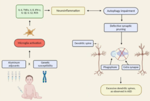
The autism spectrum disorders (ASD) are a group of related neurodevelopmental disorders that have been increasing in incidence since the 1980s. Despite a considerable amount of data being collected from cases, a central mechanism has not been offered. A careful review of ASD cases discloses a number of events that adhere to an immunoexcitotoxic mechanism. This mechanism explains the link between excessive vaccination, use of aluminum and ethylmercury as vaccine adjuvants, food allergies, gut dysbiosis, and abnormal formation of the developing brain. It has now been shown that chronic microglial activation is present in autistic brains from age 5 years to age 44 years. A considerable amount of evidence, both experimental and clinical, indicates that repeated microglial activation can initiate priming of the microglia and that subsequent stimulation can produce an exaggerated microglial response that can be prolonged. It is also known that one phenotypic form of microglia activation can result in an outpouring of neurotoxic levels of the excitotoxins, glutamate and quinolinic acid. Studies have shown that careful control of brain glutamate levels is essential to brain pathway development and that excesses can result in arrest of neural migration, as well as dendritic and synaptic loss. It has also been shown that certain cytokines, such as TNF-alpha, can, via its receptor, interact with glutamate receptors to enhance the neurotoxic reaction. To describe this interaction I have coined the term immunoexcitotoxicity, which is described in this article.
