Abstract

Mercury toxicity mediated by different forms of mercury is a major health problem; however, the molecular mechanisms underlying toxicity remain elusive. We analyzed the effects of mercuric chloride (HgCl(2)) and monomethylmercury (MeHg) on the proteins of the mammalian thioredoxin system, thioredoxin reductase (TrxR) and thioredoxin (Trx), and of the glutaredoxin system, glutathione reductase (GR) and glutaredoxin (Grx). HgCl(2) and MeHg inhibited recombinant rat TrxR with IC(50) values of 7.2 and 19.7 nm, respectively. Fully reduced human Trx1 bound mercury and lost all five free thiols and activity after incubation with HgCl(2) or MeHg, but only HgCl(2) generated dimers. Mass spectra analysis demonstrated binding of 2.5 mol of Hg(2+) and 5 mol of MeHg(+)/mol of Trx1 with the very strong Hg(2+) complexes involving active site and structural disulfides. Inhibition of both TrxR and Trx activity was observed in HeLa and HEK 293 cells treated with HgCl(2) or MeHg. GR was inhibited by HgCl(2) and MeHg in vitro, but no decrease in GR activity was detected in cell extracts treated with mercurials. Human Grx1 showed similar reactivity as Trx1 with both mercurial compounds, with the loss of all free thiols and Grx dimerization in the presence of HgCl(2), but no inhibition of Grx activity was observed in lysates of HeLa cells exposed to mercury. Overall, mercury inhibition was selective toward the thioredoxin system. In particular, the remarkable potency of the mercury compounds to bind to the selenol-thiol in the active site of TrxR should be a major molecular mechanism of mercury toxicity.

More
  • March 4, 2008

Excerpt:
“Exposure to environmental toxins is the likely etiology for MtD in autism. This dysfunction then contributes to a number of diagnostic symptoms and comorbidities observed in autism including: cognitive impairment, language deficits, abnormal energy metabolism, chronic gastrointestinal problems, abnormalities in fatty acid oxidation, and increased oxidative stress. MtD and oxidative stress may also explain the high male to female ratio found in autism due to increased male vulnerability to these dysfunctions.”

More
  • February 21, 2008

Excerpt:
“In autism, over-zealous neuroinflammatory responses could not only influence neural developmental processes, but may more significantly impair neural signaling involved in cognition in an ongoing fashion.”

More
  • February 21, 2008

Abstract

This study should be viewed as hypothesis-generating – a first step in examining the potential role of environmental mercury and childhood developmental disorders. Nothing is known about specific exposure routes, dosage, timing, and individual susceptibility. We suspect that persistent low-dose exposures to various environmental toxicants, including mercury, that occur during critical windows of neural development among genetically susceptible children (with a diminished capacity for metabolizing accumulated toxicants) may increase the risk for developmental disorders such as autism. Successfully identifying the specific combination of environmental exposures and genetic susceptibilities can inform the development of targeted prevention intervention strategies.

More
  • February 12, 2008

Exceprts:
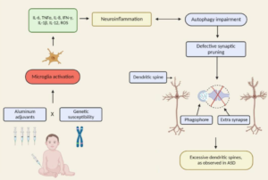
We also discuss evidence implicating oxidative stress, neuroglial activation and neuroimmunity in autism.

“Oxidative stress is another possible cause of Purkinje cell loss and other neuroanatomical changes described in autistic brains (reviewed in (37, 113)). Oxidative stress occurs when the levels of reactive oxygen species exceed the antioxidant capacities of a cell, often leading to cell death. Because of its very high oxygen demands and limited anti-oxidant capacity, the brain is thought to be relatively vulnerable to oxidative stress (111). Several studies have shown decreased levels of antioxidants such as superoxide dismutase, transferrin and ceruloplasmin in the blood or serum of patients with ASD (38, 108, 222). Significant elevations in biomarker profiles indicating increased oxidative stress, such as increased lipid peroxidation, have also been documented in autism (38, 107, 229).Interestingly, in one report the alterations in antioxidant proteins were linked specifically to regressive autism, suggesting a postnatal environmental effect (38). Polymorphisms in metabolic pathway genes may contribute to the increased oxidative stress in autism (108). Advanced glycationend products have also been reported to be elevated in both the brain tissue and serum of autistic patients, a change which can also lead to increased oxidative damage (23,110).”

More
  • October 17, 2007

Abstract

The objective of this study was to estimate the prevalence of autistic spectrum disorder (ASD) and identify its clinical characterization, and medical conditions in a paediatric population in Portugal. A school survey was conducted in elementary schools, targeting 332 808 school-aged children in the mainland and 10 910 in the Azores islands. Referred children were directly assessed using the Diagnostic and Statistical Manual of Mental Disorders (4th edn), the Autism Diagnostic Interview–Revised, and the Childhood Autism Rating Scale. Clinical history and a laboratory investigation was performed. In parallel, a systematic multi-source search of children known to have autism was carried out in a restricted region. The global prevalence of ASD per 10 000 was 9.2 in mainland, and 15.6 in the Azores, with intriguing regional differences. A diversity of associated medical conditions was documented in 20%, with an unexpectedly high rate of mitochondrial respiratory chain disorders.

More
  • September 18, 2007

Abstract

Autistic disorder is a heterogeneous disorder. The majority of the cases are idiopathic, and only a small number of the autistic children have associated secondary diagnosis. This article reports 2 children with mitochondrial disorders associated with autistic disorder fulfilling the diagnostic criteria of the American Psychiatric Association Manual of Psychiatric Diseases, 4th edition, and briefly reviews the literature on autistic disorder associated with mitochondrial disorders.

More
  • September 1, 2007

Excerpt:

“Elevation of cerebrospinal fluid levels of tumor necrosis factor-alpha was significantly higher (mean = 104.10 pg/mL) than concurrent serum levels (mean = 2.78 pg/mL) in all of the patients studied. The ratio of the cerebrospinal fluid levels to serum levels averaged 53.7:1. This ratio is significantly higher than the elevations reported for other pathological states for which cerebrospinal fluid and serum tumor necrosis factor-alpha levels have been simultaneously measured.”

More
  • June 1, 2007

Excerpt:

“There was a significant dose-response relationship between the severity of the regressive ASDs observed and the total mercury dose children received from Thimerosal-containing vaccines/Rho (D)-immune globulin preparations. Based upon differential diagnoses, 8 of 9 patients examined were exposed to significant mercury from Thimerosal-containing biologic/vaccine preparations during their fetal/infant developmental periods, and subsequently, between 12 and 24 mo of age, these previously normally developing children suffered mercury toxic encephalopathies that manifested with clinical symptoms consistent with regressive ASDs. Evidence for mercury intoxication should be considered in the differential diagnosis as contributing to some regressive ASDs.”

More
  • May 15, 2007