20 Papers

Some have asked for a smaller set of papers in order to get a more digestible overview of vaccine autism causation. Here we present 20 reviews toward that end.

Excerpts:
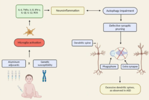
“As a result of these pieces of evidence (epidemiological, clinical and preclinical data) pointing to a potential causal association between early ABA (aluminum-based adjuvants) exposure and increased ASD risk, new hypotheses regarding neurological and immunological consequences of ABA-containing vaccines and novel clinical strategies (i.e., postponing of ABA-containing vaccines and replacement of ABAs with calcium phosphate are now being considered.“

“Our review presents the lack of fundamental scientific data demonstrating that Al adjuvants are safe and do not induce any long-term side effects. It also supports further investigation related to the effects of early Al adjuvant exposures occurring in combination with genetic susceptibility factors, including autophagy, immune and inflammation process genes. As accumulating evidence shows that modulating the levels of autophagy may increase the risk of NDDs, such studies will elucidate a new etiology for these complex disorders and contribute to develop potential new diagnostic and therapeutic tools.”
More
  • August 31, 2022

Except:
“There is a growing body of work to support the role of inflammatory cytokines in ASD. An emerging focus of research into the etiology of ASD has suggested neuroinflammation as one of the major candidates underlying the biologica model [5]. Plasma levels of IL-1β, IL-6 and IL-8 were increased in children with ASD and correlated with regressive autism, as well as impaired communication and aberrant behavior [6-8]. Vargas [9] showed an active neuroinflammatory process in the cerebral cortex, white matter, and in the cerebellum of autistic patients. Immunocytochemichal studies showed marked activation of microglia [5].”

More
  • January 1, 2019

Excerpts:

“The likelihood of the child having ASD more than doubled among children with food allergy compared with those without food allergy; children with respiratory and skin allergy were also significantly more likely to have ASD, but at a lesser magnitude. While no sex difference was found for food allergy, boys with ASD were significantly more likely than girls with ASD to have respiratory and skin allergy.”

” It may be that GI dysfunction, seizures, and sleep disorder, in addition to food, respiratory, and skin allergies, are medical comorbidities that characterize the immune-mediated subtype of ASD.”

“In the Discussion section of their article, Xu and colleagues review other aspects of immune dysfunction reported in ASD, including abnormalities in peripheral immunoglobulins, imbalance of T-cell subsets, and increased levels of proinflammatory cytokines in postmortem brains of patients with ASD. Considering the significant association between food, respiratory, and skin allergy in children with ASD reported by Xu and colleagues, in conjunction with numerous studies documenting aspects of immune dysfunction in patients with ASD and specific animal models of ASD, evidence continues to mount that an immune-mediated subtype of ASD should continue to be pursued and defined.”

More
  • June 8, 2018

Excerpt:
“The literature strongly supports that autism is most accurately seen as an acquired cellular detoxification deficiency syndrome with heterogeneous genetic predisposition that manifests pathophysiologic consequences of accumulated, run-away cellular toxicity. At a more general level, it is a form of a toxicant-induced loss of tolerance of toxins, and of chronic and sustained ER overload (“ER hyperstress”), contributing to neuronal and glial apoptosis via the unfolded protein response (UPR). Inherited risk of impaired cellular detoxification and circulating metal re-toxification in neurons and glial cells accompanied by chronic UPR is key.”

More
  • March 16, 2018

Conclusions
Results of the current meta-analysis revealed that mercury is an important causal factor in the etiology of ASD. It seems that the detoxification and excretory mechanisms are impaired in ASD patients which lead to accumulation of mercury in the body. Future additional studies on mercury levels in different tissues of ASD patients should be undertaken.

More
  • September 4, 2017

Excerpt:
“Environmental mercury is neurotoxic at doses well below the current reference levels considered to be safe, with evidence of neurotoxicity in children exposed to environmental sources including fish consumption and ethylmercury-containing vaccines. Possible neurotoxic mechanisms of mercury include direct effects on sulfhydryl groups, pericytes and cerebral endothelial cells, accumulation within astrocytes, microglial activation, induction of chronic oxidative stress, activation of immune-inflammatory pathways and impairment of mitochondrial functioning. (Epi-)genetic factors which may increase susceptibility to the toxic effects of mercury in ASD include the following: a greater propensity of males to the long-term neurotoxic effects of postnatal exposure and genetic polymorphisms in glutathione transferases and other glutathione-related genes and in selenoproteins. Furthermore, immune and inflammatory responses to immunisations with mercury-containing adjuvants are strongly influenced by polymorphisms in the human leukocyte antigen (HLA) region and by genes encoding effector proteins such as cytokines and pattern recognition receptors. Some epidemiological studies investigating a possible relationship between high environmental exposure to methylmercury and impaired neurodevelopment have reported a positive dose-dependent effect.”

More
  • July 22, 2017

Excerpts:
“The difference in manifested toxicity of MeHg and EtHg are likely the result of the differences in exposure, metabolism, and elimination from the body, rather than differences in mechanisms of action between the two.”

“Summary and Conclusions

There are many commonalities/similarities in the mechanisms of toxic action of methylmercury and ethylmercury (from thimerosal)… Evidence for the similarity of the various mechanisms of toxicity include the following:

• Both MeHg and EtHg bind to the amino acid cysteine (Clarkson 1995; Wu et al. 2008)…

• Both decrease glutathione activity, thus providing less protection from the oxidative stress caused by MeHg and EtHg (Carocci et al. 2014; Ndountse and Chan (2008); Choi et al. 1996; Franco et al. 2006; Mori et al. 2007; Muller et al. 2001; Ndountse and Chan 2008; Wu et al. 2008)…

• Both disrupt glutamate homeostasis (Farina et al. 2003a, b; Manfroi et al. 2004; Mutkus et al. 2005; Yin et al. 2007).

• Both cause oxidative stress/creation of ROS (Dreiem and Seegal 2007; Garg and Chang 2006; Myhre et al. 2003; Sharpe et al. 2012; Yin et al. 2007)…

• Both cause effects on receptor binding/neurotransmitter release involving one or more transmitters (Basu et al. 2008; Coccini et al. 2000; Cooper et al. 2003; Fonfria et al. 2001; Ida-Eto et al. 2011; Ndountse and Chan 2008; Yuan and Atchison 2003).

• Both cause DNA damage or impair DNA synthesis (Burke et al. 2006; Sharpe et al. 2012; Wu et al. 2008).”

More
  • January 1, 2016

Conclusion
There is now sufficient evidence from both human and animal studies showing that cumulative exposure to aluminium adjuvants is not as benign as previously assumed. Given that vaccines are the only medical intervention that we attempt to deliver to every living human on earth and that by far the largest target population for vaccination are healthy children, a better appreciation and understanding of vaccine adjuvant risks appears warranted.

More
  • June 10, 2014