Antioxidant

A substance that opposes oxidation or prevents or makes difficult reactions made easier by oxygen

Highlights

• Thimerosal (TM) significantly affects mitochondrial bioenergetics in the brain.
• Mitochondrial integrity (membrane potential) was maintained after acute TM treatment.
• Ethylmercury released after the breakdown of TM compromised the cholinergic system.
• The brain is more sensitive to oxidative stress induced by TM compared to the liver.

More
  • February 6, 2024

Excerpt:
“Accumulating evidence implies the gut-brain axis as a pathway for MeHg harmful neurotoxic effects and a potential factor for later neurodegenerative disorders. The MeHg may induce a hormesis-related neuronal toxicity. Hormesis is an important redox dependent aging-associated neurodegenerative/ neuroprotective issue (Calabrese et al., 2010). The use of antioxidants, such as plant polyphenols (Calabrese et al., 2010; Leri et al., 2020) and protective nutrients (Oria et al., 2020) may be beneficial in reducing the MeHg-driven neuroinflammatory state and associated cell death with the interplay of the intestinal microbiota.”

More
  • November 3, 2020

Excerpt:
“In this review article, we examine the connections between early disruption of the developing microbiome and gastrointestinal tract function, with particular regard to susceptibility to autism. The biological mechanisms that accompany individuals with autism are reviewed in this manuscript including immune system dysregulation, inflammation, oxidative stress, metabolic and methylation abnormalities as well as gastrointestinal distress.”

More
  • August 15, 2018

Abstract

Mercury toxicity is a highly interesting topic in biomedicine due to the severe endpoints and treatment limitations. Selenite serves as an antagonist of mercury toxicity, but the molecular mechanism of detoxification is not clear. Inhibition of the selenoenzyme thioredoxin reductase (TrxR) is a suggested mechanism of toxicity. Here, we demonstrated enhanced inhibition of activity by inorganic and organic mercury compounds in NADPH-reduced TrxR, consistent with binding of mercury also to the active site selenolthiol. On treatment with 5 μM selenite and NADPH, TrxR inactivated by HgCl(2) displayed almost full recovery of activity. Structural analysis indicated that mercury was complexed with TrxR, but enzyme-generated selenide removed mercury as mercury selenide, regenerating the active site selenocysteine and cysteine residues required for activity. The antagonistic effects on TrxR inhibition were extended to endogenous antioxidants, such as GSH, and clinically used exogenous chelating agents BAL, DMPS, DMSA, and α-lipoic acid. Consistent with the in vitro results, recovery of TrxR activity and cell viability by selenite was observed in HgCl(2)-treated HEK 293 cells. These results stress the role of TrxR as a target of mercurials and provide the mechanism of selenite as a detoxification agent for mercury poisoning.

More
  • September 1, 2010

Excerpt:
“Exposure to oxidative stress via the sulfhydryl reagent thimerosal resulted in a greater decrease in the GSH/GSSG ratio and increase in free radical generation in autism compared to control cells. Acute exposure to physiological levels of nitric oxide decreased mitochondrial membrane potential to a greater extent in the autism LCLs, although GSH/GSSG and ATP concentrations were similarly decreased in both cell lines. These results suggest that the autism LCLs exhibit a reduced glutathione reserve capacity in both cytosol and mitochondria that may compromise antioxidant defense and detoxification capacity under prooxidant conditions.”

More
  • March 23, 2009

Shows a potential link between mercury and the autopsied brains of young people with autism. A marker for oxidative stress was 68.9% higher in autistic brain issue than controls (a statistically significant result), while mercury levels were 68.2% higher.

Excerpt:
“The preliminary data suggest a need for more extensive studies of oxidative stress, its relationship to the environmental factors and its possible attenuation by antioxidants in autism.”

More
  • June 30, 2008

Exceprts:
We also discuss evidence implicating oxidative stress, neuroglial activation and neuroimmunity in autism.

“Oxidative stress is another possible cause of Purkinje cell loss and other neuroanatomical changes described in autistic brains (reviewed in (37, 113)). Oxidative stress occurs when the levels of reactive oxygen species exceed the antioxidant capacities of a cell, often leading to cell death. Because of its very high oxygen demands and limited anti-oxidant capacity, the brain is thought to be relatively vulnerable to oxidative stress (111). Several studies have shown decreased levels of antioxidants such as superoxide dismutase, transferrin and ceruloplasmin in the blood or serum of patients with ASD (38, 108, 222). Significant elevations in biomarker profiles indicating increased oxidative stress, such as increased lipid peroxidation, have also been documented in autism (38, 107, 229).Interestingly, in one report the alterations in antioxidant proteins were linked specifically to regressive autism, suggesting a postnatal environmental effect (38). Polymorphisms in metabolic pathway genes may contribute to the increased oxidative stress in autism (108). Advanced glycationend products have also been reported to be elevated in both the brain tissue and serum of autistic patients, a change which can also lead to increased oxidative damage (23,110).”

More
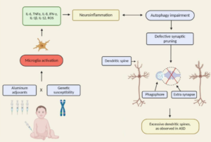
  • October 17, 2007

Excerpt:
“The metabolic results indicated that plasma methionine and the ratio of S-adenosylmethionine (SAM) to S-adenosylhomocysteine (SAH), an indicator of methylation capacity, were significantly decreased in the autistic children relative to age-matched controls. In addition, plasma levels of cysteine, glutathione, and the ratio of reduced to oxidized glutathione, an indication of antioxidant capacity and redox homeostasis, were significantly decreased. Differences in allele frequency and/or significant gene-gene interactions were found for relevant genes encoding the reduced folate carrier (RFC 80G > A), transcobalamin II (TCN2 776G > C), catechol-O-methyltransferase (COMT 472G > A), methylenetetrahydrofolate reductase (MTHFR 677C > T and 1298A > C), and glutathione-S-transferase (GST M1). We propose that an increased vulnerability to oxidative stress (endogenous or environmental) may contribute to the development and clinical manifestations of autism.”

More
  • December 17, 2006