Abstract
There is a compelling argument that the occurrence of regressive autism is attributable to genetic and chromosomal abnormalities, arising from the overuse of vaccines, which subsequently affects the stability and function of the autonomic nervous system and physiological systems. That sense perception is linked to the autonomic nervous system and the function of the physiological systems enables us to examine the significance of autistic symptoms from a systemic perspective. Failure of the excretory system influences elimination of heavy metals and facilitates their accumulation and subsequent manifestation as neurotoxins: the long-term consequences of which would lead to neurodegeneration, cognitive and developmental problems. It may also influence regulation of neural hyperthermia. This article explores the issues and concludes that sensory dysfunction and systemic failure, manifested as autism, is the inevitable consequence arising from subtle DNA alteration and consequently from the overuse of vaccines.
Genes
Excerpts:
“Such special cases suggest that the pathophysiology of autism may comprise pathways that are directly or indirectly involved in mitochondrial energy production…”
Excerpt:
“Depletion of intracellular GSH with buthionine sulfoximine treatment greatly increased the K562 cell growth inhibitory effects of thimerosal, which showed that intracellular glutathione had a major role in protecting cells from thimerosal. “
Excerpt:
“The metabolic results indicated that plasma methionine and the ratio of S-adenosylmethionine (SAM) to S-adenosylhomocysteine (SAH), an indicator of methylation capacity, were significantly decreased in the autistic children relative to age-matched controls. In addition, plasma levels of cysteine, glutathione, and the ratio of reduced to oxidized glutathione, an indication of antioxidant capacity and redox homeostasis, were significantly decreased. Differences in allele frequency and/or significant gene-gene interactions were found for relevant genes encoding the reduced folate carrier (RFC 80G > A), transcobalamin II (TCN2 776G > C), catechol-O-methyltransferase (COMT 472G > A), methylenetetrahydrofolate reductase (MTHFR 677C > T and 1298A > C), and glutathione-S-transferase (GST M1). We propose that an increased vulnerability to oxidative stress (endogenous or environmental) may contribute to the development and clinical manifestations of autism.”
Abstract
Autism is defined behaviorally, as a syndrome of abnormalities involving language, social reciprocity and hyperfocus or reduced behavioral flexibility. It is clearly heterogeneous, and it can be accompanied by unusual talents as well as by impairments, but its underlying biological and genetic basis in unknown. Autism has been modeled as a brain-based, strongly genetic disorder, but emerging findings and hypotheses support a broader model of the condition as a genetically influenced and systemic. These include imaging, neuropathology and psychological evidence of pervasive (and not just specific) brain and phenotypic features; postnatal evolution and chronic persistence of brain, behavior and tissue changes (e.g. inflammation) and physical illness symptomatology (e.g. gastrointestinal, immune, recurrent infection); overlap with other disorders; and reports of rate increases and improvement or recovery that support a role for modulation of the condition by environmental factors (e.g. exacerbation or triggering by toxins, infectious agents, or others stressors, or improvement by treatment). Modeling autism more broadly encompasses previous work, but also encourages the expansion of research and treatment to include intermediary domains of molecular and cellular mechanisms, as well as chronic tissue, metabolic and somatic changes previously addressed only to a limited degree. The heterogeneous biologies underlying autism may conceivably converge onto the autism profile via multiple mechanisms on the one hand and processing and connectivity abnormalities on the other may illuminate relevant final common pathways and contribute to focusing on the search for treatment targets in this biologically and etiologically heterogeneous behavioral syndrome.
Excerpt:
“Recently, it was found that autistic children had a higher mercury exposure during pregnancy due to maternal dental amalgam and thimerosal-containing immunoglobulin shots. It was hypothesized that children with autism have a decreased detoxification capacity due to genetic polymorphism. In vitro, mercury and thimerosal in levels found several days after vaccination inhibit methionine synthetase (MS) by 50%. Normal function of MS is crucial in biochemical steps necessary for brain development, attention and production of glutathione, an important antioxidative and detoxifying agent. Repetitive doses of thimerosal leads to neurobehavioral deteriorations in autoimmune susceptible mice, increased oxidative stress and decreased intracellular levels of glutathione in vitro. Subsequently, autistic children have significantly decreased level of reduced glutathione. Promising treatments of autism involve detoxification of mercury, and supplementation of deficient metabolites.”
Excerpt:
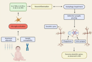
“The promoters of genes up-regulated by aluminum are enriched in binding sites for the stress-inducible transcription factors HIF-1 and NF-kappaB, suggesting a role for aluminum, HIF-1 and NF-kappaB in driving atypical, pro-inflammatory and pro-apoptotic gene expression. The effect of aluminum on specific stress-related gene expression patterns in human brain cells clearly warrant further investigation.”
Excerpts:
“However, testosterone which appeared protective at very low levels (0.01 to 0.1 micromolar), dramatically increased neuron death at higher levels (0.5 to 1.0 micromolar). In fact, 1.0 micromolar levels of testosterone that by itself did not significantly increase neuron death (red flattened oval), within 3 hours when added with 50 nanomolar thimerosal (solid circles) caused 100% neuron death.”
“These testosterone results, while not conclusive because of the in vitro neuron culture type of testing, clearly demonstrated that male versus female hormones may play a major role in autism risk and may explain the high ratio of boys to girls in autism (4 to 1) and autism related disorders.“
Abstract
Methylation events play a critical role in the ability of growth factors to promote normal development. Neurodevelopmental toxins, such as ethanol and heavy metals, interrupt growth factor signaling, raising the possibility that they might exert adverse effects on methylation. We found that insulin-like growth factor-1 (IGF-1)- and dopamine-stimulated methionine synthase (MS) activity and folate-dependent methylation of phospholipids in SH-SY5Y human neuroblastoma cells, via a PI3-kinase- and MAP-kinase-dependent mechanism. The stimulation of this pathway increased DNA methylation, while its inhibition increased methylation-sensitive gene expression. Ethanol potently interfered with IGF-1 activation of MS and blocked its effect on DNA methylation, whereas it did not inhibit the effects of dopamine. Metal ions potently affected IGF-1 and dopamine-stimulated MS activity, as well as folate-dependent phospholipid methylation: Cu(2+) promoted enzyme activity and methylation, while Cu(+), Pb(2+), Hg(2+) and Al(3+) were inhibitory. The ethylmercury-containing preservative thimerosal inhibited both IGF-1- and dopamine-stimulated methylation with an IC(50) of 1 nM and eliminated MS activity. Our findings outline a novel growth factor signaling pathway that regulates MS activity and thereby modulates methylation reactions, including DNA methylation. The potent inhibition of this pathway by ethanol, lead, mercury, aluminum and thimerosal suggests that it may be an important target of neurodevelopmental toxins.
Conclusions. Autoimmunity was increased significantly in families with PDD compared with those of healthy and autoimmune control subjects.
