Thimerosal

Abstract

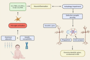
The autism spectrum disorders (ASD) are a group of related neurodevelopmental disorders that have been increasing in incidence since the 1980s. Despite a considerable amount of data being collected from cases, a central mechanism has not been offered. A careful review of ASD cases discloses a number of events that adhere to an immunoexcitotoxic mechanism. This mechanism explains the link between excessive vaccination, use of aluminum and ethylmercury as vaccine adjuvants, food allergies, gut dysbiosis, and abnormal formation of the developing brain. It has now been shown that chronic microglial activation is present in autistic brains from age 5 years to age 44 years. A considerable amount of evidence, both experimental and clinical, indicates that repeated microglial activation can initiate priming of the microglia and that subsequent stimulation can produce an exaggerated microglial response that can be prolonged. It is also known that one phenotypic form of microglia activation can result in an outpouring of neurotoxic levels of the excitotoxins, glutamate and quinolinic acid. Studies have shown that careful control of brain glutamate levels is essential to brain pathway development and that excesses can result in arrest of neural migration, as well as dendritic and synaptic loss. It has also been shown that certain cytokines, such as TNF-alpha, can, via its receptor, interact with glutamate receptors to enhance the neurotoxic reaction. To describe this interaction I have coined the term immunoexcitotoxicity, which is described in this article.
More
  • November 1, 2014

Abstract
Reported rates of autism have increased sharply in the United States and the United Kingdom. One possible factor underlying these increases is increased exposure to mercury through thimerosal-containing vaccines, but vaccine exposures need to be evaluated in the context of cumulative exposures during gestation and early infancy. Differential rates of postnatal mercury elimination may explain why similar gestational and infant exposures produce variable neurological effects. First baby haircut samples were obtained from 94 children diagnosed with autism using Diagnostic and Statistical Manual of Mental Disorders, 4th edition (DSM IV) criteria and 45 age- and gender-matched controls. Information on diet, dental amalgam fillings, vaccine history, Rho D immunoglobulin administration, and autism symptom severity was collected through a maternal survey questionnaire and clinical observation. Hair mercury levels in the autistic group were 0.47 ppm versus 3.63 ppm in controls, a significant difference. The mothers in the autistic group had significantly higher levels of mercury exposure through Rho D immunoglobulin injections and amalgam fillings than control mothers. Within the autistic group, hair mercury levels varied significantly across mildly, moderately, and severely autistic children, with mean group levels of 0.79, 0.46, and 0.21 ppm, respectively. Hair mercury levels among controls were significantly correlated with the number of the mothers’ amalgam fillings and their fish consumption as well as exposure to mercury through childhood vaccines, correlations that were absent in the autistic group. Hair excretion patterns among autistic infants were significantly reduced relative to control. These data cast doubt on the efficacy of traditional hair analysis as a measure of total mercury exposure in a subset of the population. In light of the biological plausibility of mercury’s role in neurodevelopmental disorders, the present study provides further insight into one possible mechanism by which early mercury exposures could increase the risk of autism.

More
  • September 4, 2014

Abstract

Autism spectrum disorders (ASDs) are complex, heterogeneous disorders caused by an interaction between genetic vulnerability and environmental factors. In an effort to better target the underlying roots of ASD for diagnosis and treatment, efforts to identify reliable biomarkers in genetics, neuroimaging, gene expression, and measures of the body’s metabolism are growing. For this article, we review the published studies of potential biomarkers in autism and conclude that while there is increasing promise of finding biomarkers that can help us target treatment, there are none with enough evidence to support routine clinical use unless medical illness is suspected. Promising biomarkers include those for mitochondrial function, oxidative stress, and immune function. Genetic clusters are also suggesting the potential for useful biomarkers.

Excerpt: “A recent review assessed the research on physiological abnormalities associated with ASD (44). The authors identified four main mechanisms that have been increasingly studied during the past decade: immunologic/inflammation, oxidative stress, environmental toxicants, and mitochondrial abnormalities. In addition, there is accumulating research on the lipid, GI systems, microglial activation, and the microbiome, and how these can also contribute to generating biomarkers associated with ASD (45, 46).

Pathways are interconnected with a defect in one likely leading to dysfunction in others. Many metabolic disorders can lead to endpoints such as impaired methylation, sulfuration, and detoxification pathways and nutritional deficiencies. Mitochondrial dysfunction, environmental risk factors, metabolic imbalances, and genetic susceptibility can all lead to oxidative stress (47), which in turn leads to inflammation, damaged cell membranes, autoimmunity (48), impaired methylation (49), cell death (48), and neurological deficits (50). The brain is highly vulnerable to oxidative stress (51), particularly in children (52) during the early part of development (47). As environmental events and metabolic imbalances affect oxidative stress and methylation, they also can affect the expression of genes.”

More
  • August 12, 2014

Excerpt:
“Intriguingly, the elevation of anterior pituitary secreting hormones occurred exclusively in male but not in female thimerosal-treated mice, demonstrating for the first time the gender bias of thimerosal-mercury toxicity with regard to endocrine system. Our results indicate that higher dose of neonatal thimerosal-mercury (20× higher than that used in human) is capable of inducing long-lasting substantial dysregulation of neurodevelopment, synaptic function, and endocrine system, which could be the causal involvements of autistic-like behavior in mice.”

More
  • March 27, 2014

CONCLUSIONS:
The negative adverse consequences on neurodevelopment observed in the present study are consistent with previous studies; this study raised serious concerns about adverse neurodevelopmental disorder such as autism in humans following the ongoing worldwide routine administration of thimerosal containing vaccines to infants.

More
  • November 15, 2013

Abstract
Thimerosal generates ethylmercury in aqueous solution and is widely used as preservative. We have investigated the toxicology of Thimerosal in normal human astrocytes, paying particular attention to mitochondrial function and the generation of specific oxidants. We find that ethylmercury not only inhibits mitochondrial respiration leading to a drop in the steady state membrane potential, but also concurrent with these phenomena increases the formation of superoxide, hydrogen peroxide, and Fenton/Haber-Weiss generated hydroxyl radical. These oxidants increase the levels of cellular aldehyde/ketones. Additionally, we find a five-fold increase in the levels of oxidant damaged mitochondrial DNA bases and increases in the levels of mtDNA nicks and blunt-ended breaks. Highly damaged mitochondria are characterized by having very low membrane potentials, increased superoxide/hydrogen peroxide production, and extensively damaged mtDNA and proteins. These mitochondria appear to have undergone a permeability transition, an observation supported by the five-fold increase in Caspase-3 activity observed after Thimerosal treatment.

More
  • June 28, 2012

Excerpts:
“Marie Curie Chairs Program, Department of Pharmacology and Physiology of Nervous System, Institute of Psychiatry and Neurology, 02-957, Warsaw, Poland.

Abstract
Thimerosal, a mercury-containing vaccine preservative, is a suspected factor in the etiology of neurodevelopmental disorders. We previously showed that its administration to infant rats causes behavioral, neurochemical and neuropathological abnormalities similar to those present in autism.”

“Since excessive accumulation of extracellular glutamate is linked with excitotoxicity, our data imply that neonatal exposure to thimerosal-containing vaccines might induce excitotoxic brain injuries, leading to neurodevelopmental disorders.”

More
  • October 21, 2011