admin

Press Release from Harvard Magazine:

“Inflammation link for autism
A neuroimaging study has shown that the brains of young men with autism spectrum disorder have low levels of translocator protein, a substance that appears to play a role in inflammation and metabolism.

This discovery by a team of HMS researchers at Massachusetts General Hospital provides an important insight into the possible origins of autism spectrum disorder.

This developmental disorder, which affects one in fifty-nine children in the United States, emerges in early childhood and is characterized by difficulty communicating and interacting with others. Although the cause is unknown, growing evidence has linked it to neuroinflammation.

One sign of neuroinflammation is elevated levels of translocator protein, which can be measured in the brain using positron-emission tomography and anatomic magnetic resonance imaging.

The research team used these imaging tools to scan the brains of fifteen young adult males with the disorder. The group included both high- and low-functioning participants with varying degrees of intellectual ability. As a control, the team scanned the brains of eighteen non-autistic young men of similar age.

The scans showed that the brains of the young men with the disorder had lower levels of the protein, compared with the brains of non-autistic participants. In fact, those participants with the most severe symptoms of the disorder tended to have the lowest expression of the protein.

The brain regions found to have low expression of the protein have previously been linked to autism spectrum disorder and are thought to govern social and cognitive capacities such as processing emotions, interpreting facial expressions, and empathy.

The researchers point out that the translocator protein has multiple complex roles, some of which promote brain health. Adequate levels of the protein are, for example, necessary for normal functioning of mitochondria. Earlier research has linked malfunctioning mitochondria in brain cells to autism spectrum disorder.

Zürcher NR et al., Molecular Psychiatry, February 2020
More
  • February 19, 2020

Excerpt:
“Results: A significant and positive correlation was found between hair metal burden (lead, aluminum, arsenic and cadmium levels) and severity of ASD symptoms (social communication deficits and repetitive, restrictive behaviors). Hair zinc level were inversely related with age while there was a negative, significant association between hair zinc level and severity of autistic symptoms (defective functional play and creativity and increase of stereotyped behavior). Lead, molybdenum and manganese hair levels were inversely correlated with cognitive level (full intelligence quotient) in ASD individuals.

Conclusions: The present study suggests the importance to combine metallomics analysis with pertinent disease features in ASD to identify potential environmental risk factors on an individual level possibly in the early developmental period.”

More
  • January 1, 2020

Discussion: The observation of predominantly intracellular aluminium in these tissues was novel and something similar has only previously been observed in cases of autism. The results suggest a strong inflammatory component in this case and support a role for aluminium in this rare and unusual case of CAA.

More
  • April 24, 2019

Except:
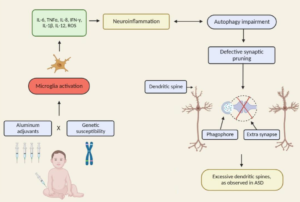
“There is a growing body of work to support the role of inflammatory cytokines in ASD. An emerging focus of research into the etiology of ASD has suggested neuroinflammation as one of the major candidates underlying the biologica model [5]. Plasma levels of IL-1β, IL-6 and IL-8 were increased in children with ASD and correlated with regressive autism, as well as impaired communication and aberrant behavior [6-8]. Vargas [9] showed an active neuroinflammatory process in the cerebral cortex, white matter, and in the cerebellum of autistic patients. Immunocytochemichal studies showed marked activation of microglia [5].”

More
  • January 1, 2019

Excerpt:

“Increased extra-axial cerebrospinal fluid (EA-CSF) have been observed in imaging studies of infant brains, who go on to develop autism. Folate deficiency can cause defects in neural development that can affect CSF production and drainage. Folate receptor alpha antibodies (FRAA) are observed in 75% of autism patients. Maternal FRAA have also been observed in the case of neural tube defects.

Folate deficiency can cause aluminum accumulation in the brain. Autistic brains have been shown to accumulate aluminum. FRAA in the child or mother can therefore explain all the observations”

More
  • November 30, 2018

Excerpt:
“This finding suggests that clinical events concerning neonatal IL-4 over-exposure, including neonatal hepatitis B vaccination and allergic asthma in human infants, may have adverse implications for brain development and cognition.”

More
  • October 22, 2018

Excerpts:

“Herein, we will discuss the accumulating literature for ASD, giving special attention to the relevant aspects of factors that may be related to the neuroimmune interface in the development of ASD, including changes in neuroplasticity.”

Commentary on the article:

“The authors rightly highlight the newest challenging frontier of autism research: the neuroimmune axis alterations. These alterations are first evident in the cells early responsible for immune responses, as they are the precursors for macrophages, dendritic, and microglial cells: monocytes or peripheral blood mononuclear cells (PBMCs). These cells show strong dysfunctions in ASD children and are committed to a pro-inflammatory state, which in turn result in long-term immune alterations (4). In ASDs, altered PBMCs are responsible for elevated pro-inflammatory cytokine production. The up-regulation of inflammatory cytokines is also reflected in brain centers of autistic patients (5): the consequences are the induction of blood–brain barrier (the immunological interface between peripheral immune system and central nervous system) disruption. Changes in BBB permeability directly influence neural plasticity, connectivity and function, triggering impairments in social interaction, communication, and behavior (3). Immunological abnormalities also influence the gastrointestinal system and the microglial innate immune cells of the central nervous system (6). The authors also discuss the role of autoimmunity in the pathogenesis of autism. Familial or virus/bacteria-infected autoimmunity could be a risk factor for autism. Even if the exact cellular and molecular pathways responsible for the induction of neuroimmune alterations are still to be further clarify, a complex interaction among epigenetic and environmental risk factors (7) could trigger the neuroimmune abnormalities, such as abnormal neuron and glia responses.”

More
  • September 9, 2018

Excerpt:
“In this review article, we examine the connections between early disruption of the developing microbiome and gastrointestinal tract function, with particular regard to susceptibility to autism. The biological mechanisms that accompany individuals with autism are reviewed in this manuscript including immune system dysregulation, inflammation, oxidative stress, metabolic and methylation abnormalities as well as gastrointestinal distress.”

More
  • August 15, 2018

Abstract

Time trends in U.S. autism prevalence from three ongoing datasets [Individuals with Disabilities Education Act, Autism and Developmental Disabilities Monitoring Network, and California Department of Developmental Services (CDDS)] are calculated using two different methods: (1) constant-age tracking of 8 year-olds and (2) age-resolved snapshots. The data are consistent across methods in showing a strong upward trend over time. The prevalence of autism in the CDDS dataset, the longest of the three data records, increased from 0.001% in the cohort born in 1931 to 1.2% among 5 year-olds born in 2012. This increase began around ~ 1940 at a rate that has gradually accelerated over time, including notable change points around birth years 1980, 1990 and, most recently, 2007.

More
  • July 5, 2018

Excerpts:

“The likelihood of the child having ASD more than doubled among children with food allergy compared with those without food allergy; children with respiratory and skin allergy were also significantly more likely to have ASD, but at a lesser magnitude. While no sex difference was found for food allergy, boys with ASD were significantly more likely than girls with ASD to have respiratory and skin allergy.”

” It may be that GI dysfunction, seizures, and sleep disorder, in addition to food, respiratory, and skin allergies, are medical comorbidities that characterize the immune-mediated subtype of ASD.”

“In the Discussion section of their article, Xu and colleagues review other aspects of immune dysfunction reported in ASD, including abnormalities in peripheral immunoglobulins, imbalance of T-cell subsets, and increased levels of proinflammatory cytokines in postmortem brains of patients with ASD. Considering the significant association between food, respiratory, and skin allergy in children with ASD reported by Xu and colleagues, in conjunction with numerous studies documenting aspects of immune dysfunction in patients with ASD and specific animal models of ASD, evidence continues to mount that an immune-mediated subtype of ASD should continue to be pursued and defined.”

More
  • June 8, 2018