Inflammation

Pathological process characterized by injury or destruction of tissues caused by a variety of cytologic and chemical reactions; usually manifested by typical signs of pain, heat, redness, swelling, and loss of function. – CRISP Thesaurus, National Institutes of Health

Excerpts:

“We found a functional architecture along the GBA that correlates with heterogeneity of ASD phenotypes, and it is characterized by ASD-associated amino acid, carbohydrate and lipid profiles predominantly encoded by microbial species in the genera Prevotella, Bifidobacterium, Desulfovibrio and Bacteroides and correlates with brain gene expression changes, restrictive dietary patterns and pro-inflammatory cytokine profiles.”

“We also show a strong association between temporal changes in microbiome composition and ASD phenotypes. In summary, we propose a framework to leverage multi-omic datasets from well-defined cohorts and investigate how the GBA influences ASD.”
More
  • June 26, 2023

Excerpts:
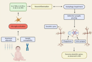
“As a result of these pieces of evidence (epidemiological, clinical and preclinical data) pointing to a potential causal association between early ABA (aluminum-based adjuvants) exposure and increased ASD risk, new hypotheses regarding neurological and immunological consequences of ABA-containing vaccines and novel clinical strategies (i.e., postponing of ABA-containing vaccines and replacement of ABAs with calcium phosphate are now being considered.“

“Our review presents the lack of fundamental scientific data demonstrating that Al adjuvants are safe and do not induce any long-term side effects. It also supports further investigation related to the effects of early Al adjuvant exposures occurring in combination with genetic susceptibility factors, including autophagy, immune and inflammation process genes. As accumulating evidence shows that modulating the levels of autophagy may increase the risk of NDDs, such studies will elucidate a new etiology for these complex disorders and contribute to develop potential new diagnostic and therapeutic tools.”

More
  • August 31, 2022

Discussion: The observation of predominantly intracellular aluminium in these tissues was novel and something similar has only previously been observed in cases of autism. The results suggest a strong inflammatory component in this case and support a role for aluminium in this rare and unusual case of CAA.

More
  • April 24, 2019

Except:
“There is a growing body of work to support the role of inflammatory cytokines in ASD. An emerging focus of research into the etiology of ASD has suggested neuroinflammation as one of the major candidates underlying the biologica model [5]. Plasma levels of IL-1β, IL-6 and IL-8 were increased in children with ASD and correlated with regressive autism, as well as impaired communication and aberrant behavior [6-8]. Vargas [9] showed an active neuroinflammatory process in the cerebral cortex, white matter, and in the cerebellum of autistic patients. Immunocytochemichal studies showed marked activation of microglia [5].”

More
  • January 1, 2019

Excerpt:
“This finding suggests that clinical events concerning neonatal IL-4 over-exposure, including neonatal hepatitis B vaccination and allergic asthma in human infants, may have adverse implications for brain development and cognition.”

More
  • October 22, 2018

Excerpts:

“Herein, we will discuss the accumulating literature for ASD, giving special attention to the relevant aspects of factors that may be related to the neuroimmune interface in the development of ASD, including changes in neuroplasticity.”

Commentary on the article:

“The authors rightly highlight the newest challenging frontier of autism research: the neuroimmune axis alterations. These alterations are first evident in the cells early responsible for immune responses, as they are the precursors for macrophages, dendritic, and microglial cells: monocytes or peripheral blood mononuclear cells (PBMCs). These cells show strong dysfunctions in ASD children and are committed to a pro-inflammatory state, which in turn result in long-term immune alterations (4). In ASDs, altered PBMCs are responsible for elevated pro-inflammatory cytokine production. The up-regulation of inflammatory cytokines is also reflected in brain centers of autistic patients (5): the consequences are the induction of blood–brain barrier (the immunological interface between peripheral immune system and central nervous system) disruption. Changes in BBB permeability directly influence neural plasticity, connectivity and function, triggering impairments in social interaction, communication, and behavior (3). Immunological abnormalities also influence the gastrointestinal system and the microglial innate immune cells of the central nervous system (6). The authors also discuss the role of autoimmunity in the pathogenesis of autism. Familial or virus/bacteria-infected autoimmunity could be a risk factor for autism. Even if the exact cellular and molecular pathways responsible for the induction of neuroimmune alterations are still to be further clarify, a complex interaction among epigenetic and environmental risk factors (7) could trigger the neuroimmune abnormalities, such as abnormal neuron and glia responses.”

More
  • September 9, 2018

Excerpt:
“In this review article, we examine the connections between early disruption of the developing microbiome and gastrointestinal tract function, with particular regard to susceptibility to autism. The biological mechanisms that accompany individuals with autism are reviewed in this manuscript including immune system dysregulation, inflammation, oxidative stress, metabolic and methylation abnormalities as well as gastrointestinal distress.”

More
  • August 15, 2018

Excerpts:

“The likelihood of the child having ASD more than doubled among children with food allergy compared with those without food allergy; children with respiratory and skin allergy were also significantly more likely to have ASD, but at a lesser magnitude. While no sex difference was found for food allergy, boys with ASD were significantly more likely than girls with ASD to have respiratory and skin allergy.”

” It may be that GI dysfunction, seizures, and sleep disorder, in addition to food, respiratory, and skin allergies, are medical comorbidities that characterize the immune-mediated subtype of ASD.”

“In the Discussion section of their article, Xu and colleagues review other aspects of immune dysfunction reported in ASD, including abnormalities in peripheral immunoglobulins, imbalance of T-cell subsets, and increased levels of proinflammatory cytokines in postmortem brains of patients with ASD. Considering the significant association between food, respiratory, and skin allergy in children with ASD reported by Xu and colleagues, in conjunction with numerous studies documenting aspects of immune dysfunction in patients with ASD and specific animal models of ASD, evidence continues to mount that an immune-mediated subtype of ASD should continue to be pursued and defined.”

More
  • June 8, 2018

Excerpt:
“CONCLUSION: Among children born EP, those who had top quartile concentrations of IL-4 and/or IL-10 on postnatal days 21 and/or 28 were more likely than their peers to have low scores on components of the NEPSY-II, OWLS-II, and WIAT-III assessments, as well as identification as having an ASD.”

More
  • May 12, 2018

Abstract
Autism spectrum disorder is a neurodevelopmental disorder of unknown aetiology. It is suggested to involve both genetic susceptibility and environmental factors including in the latter environmental toxins. Human exposure to the environmental toxin aluminium has been linked, if tentatively, to autism spectrum disorder. Herein we have used transversely heated graphite furnace atomic absorption spectrometry to measure, for the first time, the aluminium content of brain tissue from donors with a diagnosis of autism. We have also used an aluminium-selective fluor to identify aluminium in brain tissue using fluorescence microscopy. The aluminium content of brain tissue in autism was consistently high. The mean (standard deviation) aluminium content across all 5 individuals for each lobe were 3.82(5.42), 2.30(2.00), 2.79(4.05) and 3.82(5.17) μg/g dry wt. for the occipital, frontal, temporal and parietal lobes respectively. These are some of the highest values for aluminium in human brain tissue yet recorded and one has to question why, for example, the aluminium content of the occipital lobe of a 15 year old boy would be 8.74 (11.59) μg/g dry wt.? Aluminium-selective fluorescence microscopy was used to identify aluminium in brain tissue in 10 donors. While aluminium was imaged associated with neurones it appeared to be present intracellularly in microglia-like cells and other inflammatory non-neuronal cells in the meninges, vasculature, grey and white matter. The pre-eminence of intracellular aluminium associated with non-neuronal cells was a standout observation in autism brain tissue and may offer clues as to both the origin of the brain aluminium as well as a putative role in autism spectrum disorder.

More
  • March 18, 2018