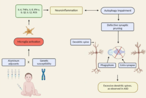
Exerpt
“any adjuvant or condition boosting immune activation of certain cells, such as Central Nervous System (CNS) microglia and macrophages, if sequentially activated by the vaccination process, whatever the adjuvant or immune stimulation principle, will trigger this mechanism, not only in the CNS, but possibly other peripheral organs and tissues containing glutamate receptors”
Neuroinflammation
Neuroinflammation is inflammation of the nervous tissue. It may be initiated in response to a variety of cues, including infection, traumatic brain injury,[1] toxic metabolites, or autoimmunity. – Molecular imaging of neuroinflammation in patients after mild traumatic brain injury: a longitudinal 123 I-CLINDE SPECT study
Excerpts:
“As a result of these pieces of evidence (epidemiological, clinical and preclinical data) pointing to a potential causal association between early ABA (aluminum-based adjuvants) exposure and increased ASD risk, new hypotheses regarding neurological and immunological consequences of ABA-containing vaccines and novel clinical strategies (i.e., postponing of ABA-containing vaccines and replacement of ABAs with calcium phosphate are now being considered.“
“Our review presents the lack of fundamental scientific data demonstrating that Al adjuvants are safe and do not induce any long-term side effects. It also supports further investigation related to the effects of early Al adjuvant exposures occurring in combination with genetic susceptibility factors, including autophagy, immune and inflammation process genes. As accumulating evidence shows that modulating the levels of autophagy may increase the risk of NDDs, such studies will elucidate a new etiology for these complex disorders and contribute to develop potential new diagnostic and therapeutic tools.”
Excerpt:
“Accumulating evidence implies the gut-brain axis as a pathway for MeHg harmful neurotoxic effects and a potential factor for later neurodegenerative disorders. The MeHg may induce a hormesis-related neuronal toxicity. Hormesis is an important redox dependent aging-associated neurodegenerative/ neuroprotective issue (Calabrese et al., 2010). The use of antioxidants, such as plant polyphenols (Calabrese et al., 2010; Leri et al., 2020) and protective nutrients (Oria et al., 2020) may be beneficial in reducing the MeHg-driven neuroinflammatory state and associated cell death with the interplay of the intestinal microbiota.”
“Background: Encephalitis, the inflammation of the brain, may be caused by an infection or an autoimmune reaction.”
“Conclusion: Gut microbiota disruption was observed in encephalitis patients, which manifested as pathogen dominance and health-promoting commensal depletion. Disease severity and brain damage may have associations with the gut microbiota or its metabolites.”
Discussion: The observation of predominantly intracellular aluminium in these tissues was novel and something similar has only previously been observed in cases of autism. The results suggest a strong inflammatory component in this case and support a role for aluminium in this rare and unusual case of CAA.
Except:
“There is a growing body of work to support the role of inflammatory cytokines in ASD. An emerging focus of research into the etiology of ASD has suggested neuroinflammation as one of the major candidates underlying the biologica model [5]. Plasma levels of IL-1β, IL-6 and IL-8 were increased in children with ASD and correlated with regressive autism, as well as impaired communication and aberrant behavior [6-8]. Vargas [9] showed an active neuroinflammatory process in the cerebral cortex, white matter, and in the cerebellum of autistic patients. Immunocytochemichal studies showed marked activation of microglia [5].”
Excerpt:
“This finding suggests that clinical events concerning neonatal IL-4 over-exposure, including neonatal hepatitis B vaccination and allergic asthma in human infants, may have adverse implications for brain development and cognition.”
Excerpts:
“Herein, we will discuss the accumulating literature for ASD, giving special attention to the relevant aspects of factors that may be related to the neuroimmune interface in the development of ASD, including changes in neuroplasticity.”
Commentary on the article:
“The authors rightly highlight the newest challenging frontier of autism research: the neuroimmune axis alterations. These alterations are first evident in the cells early responsible for immune responses, as they are the precursors for macrophages, dendritic, and microglial cells: monocytes or peripheral blood mononuclear cells (PBMCs). These cells show strong dysfunctions in ASD children and are committed to a pro-inflammatory state, which in turn result in long-term immune alterations (4). In ASDs, altered PBMCs are responsible for elevated pro-inflammatory cytokine production. The up-regulation of inflammatory cytokines is also reflected in brain centers of autistic patients (5): the consequences are the induction of blood–brain barrier (the immunological interface between peripheral immune system and central nervous system) disruption. Changes in BBB permeability directly influence neural plasticity, connectivity and function, triggering impairments in social interaction, communication, and behavior (3). Immunological abnormalities also influence the gastrointestinal system and the microglial innate immune cells of the central nervous system (6). The authors also discuss the role of autoimmunity in the pathogenesis of autism. Familial or virus/bacteria-infected autoimmunity could be a risk factor for autism. Even if the exact cellular and molecular pathways responsible for the induction of neuroimmune alterations are still to be further clarify, a complex interaction among epigenetic and environmental risk factors (7) could trigger the neuroimmune abnormalities, such as abnormal neuron and glia responses.”
Excerpt:
“The literature strongly supports that autism is most accurately seen as an acquired cellular detoxification deficiency syndrome with heterogeneous genetic predisposition that manifests pathophysiologic consequences of accumulated, run-away cellular toxicity. At a more general level, it is a form of a toxicant-induced loss of tolerance of toxins, and of chronic and sustained ER overload (“ER hyperstress”), contributing to neuronal and glial apoptosis via the unfolded protein response (UPR). Inherited risk of impaired cellular detoxification and circulating metal re-toxification in neurons and glial cells accompanied by chronic UPR is key.”
INTERPRETATION:
Our findings suggest that predisposition to autoimmunity, and immune/inflammatory activation, may be associated with autistic regression.
