Excerpt:
“CONCLUSION: Among children born EP, those who had top quartile concentrations of IL-4 and/or IL-10 on postnatal days 21 and/or 28 were more likely than their peers to have low scores on components of the NEPSY-II, OWLS-II, and WIAT-III assessments, as well as identification as having an ASD.”
Abstract
There are similarities between the immune response following immunization with aluminum adjuvants and the immune response elicited by some helminthic parasites, including stimulation of immunoglobulin E (IgE) and eosinophilia. Immunization with aluminum adjuvants, as with helminth infection, induces a Th2 type cell mediated immune response, including eosinophilia, but does not induce an environment conducive to the induction of regulatory mechanisms. Helminths play a role in what is known as the hygiene hypothesis, which proposes that decreased exposure to microbes during a critical time in early life has resulted in the increased prevalence and morbidity of asthma and atopic disorders over the past few decades, especially in Western countries. In addition, gut and lung microbiome composition and their interaction with the immune system plays an important role in a properly regulated immune system. Disturbances in microbiome composition are a risk factor for asthma and allergies. We propose that immunization with aluminum adjuvants in general is not favorable for induction of regulatory mechanisms and, in the context of the hygiene hypothesis and microbiome theory, can be viewed as an amplifying factor and significant contributing risk factor for allergic diseases, especially in a genetically susceptible subpopulation.
Abstract
Autism spectrum disorder is a neurodevelopmental disorder of unknown aetiology. It is suggested to involve both genetic susceptibility and environmental factors including in the latter environmental toxins. Human exposure to the environmental toxin aluminium has been linked, if tentatively, to autism spectrum disorder. Herein we have used transversely heated graphite furnace atomic absorption spectrometry to measure, for the first time, the aluminium content of brain tissue from donors with a diagnosis of autism. We have also used an aluminium-selective fluor to identify aluminium in brain tissue using fluorescence microscopy. The aluminium content of brain tissue in autism was consistently high. The mean (standard deviation) aluminium content across all 5 individuals for each lobe were 3.82(5.42), 2.30(2.00), 2.79(4.05) and 3.82(5.17) μg/g dry wt. for the occipital, frontal, temporal and parietal lobes respectively. These are some of the highest values for aluminium in human brain tissue yet recorded and one has to question why, for example, the aluminium content of the occipital lobe of a 15 year old boy would be 8.74 (11.59) μg/g dry wt.? Aluminium-selective fluorescence microscopy was used to identify aluminium in brain tissue in 10 donors. While aluminium was imaged associated with neurones it appeared to be present intracellularly in microglia-like cells and other inflammatory non-neuronal cells in the meninges, vasculature, grey and white matter. The pre-eminence of intracellular aluminium associated with non-neuronal cells was a standout observation in autism brain tissue and may offer clues as to both the origin of the brain aluminium as well as a putative role in autism spectrum disorder.
Excerpt:
“The literature strongly supports that autism is most accurately seen as an acquired cellular detoxification deficiency syndrome with heterogeneous genetic predisposition that manifests pathophysiologic consequences of accumulated, run-away cellular toxicity. At a more general level, it is a form of a toxicant-induced loss of tolerance of toxins, and of chronic and sustained ER overload (“ER hyperstress”), contributing to neuronal and glial apoptosis via the unfolded protein response (UPR). Inherited risk of impaired cellular detoxification and circulating metal re-toxification in neurons and glial cells accompanied by chronic UPR is key.”
“Rates of regression were much lower using retrospective or categorical measures (from 29 to 47%). Agreement among different measurement methods was low. Declining trajectories of development, consistent with a regressive onset pattern, are common in children with ASD and may be more the rule than the exception.”
“CONCLUSIONS:
the results of this preclinical study, consistent with previous studies on mice and rats, reveals that neonatal dose-dependent exposure to Thimerosal mimicking the childhood vaccine schedule can induce abnormal social interactions and stereotyped behaviors similar to those observed in neurodevelopmental disorders such as autism, and, for the first time, revealed that these abnormalities may be ameliorated by ALA. This indicates that ALA may protect against mercurial-induced abnormal behaviors.”
Abstract
Organic abnormalities with neuroinflammatory and psychiatric consequences involving abnormal kynurenine and purine metabolism, neurotransmitter and cytokine imbalances, and altered levels of nutrients and metabolites are noted in autism, and many of these abnormalities-specifically including increased intestinal permeability, microbial metabolites, and heightened serum levels of endotoxin-originate from the gut.
Excerpt:
“[S]tudies linking IL-6 and autism, suggesting potential novel therapeutic pathways for a subtype of ASD individuals. This is the first report demonstrating that glial dysfunctions could contribute to non-syndromic autism pathophysiology using iPSCs modeling disease technology.”
Conclusions
Results of the current meta-analysis revealed that mercury is an important causal factor in the etiology of ASD. It seems that the detoxification and excretory mechanisms are impaired in ASD patients which lead to accumulation of mercury in the body. Future additional studies on mercury levels in different tissues of ASD patients should be undertaken.
Abstract
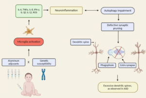
The conceptualisation of autistic spectrum disorder and Alzheimer’s disease has undergone something of a paradigm shift in recent years and rather than being viewed as single illnesses with a unitary pathogenesis and pathophysiology they are increasingly considered to be heterogeneous syndromes with a complex multifactorial aetiopathogenesis, involving a highly complex and diverse combination of genetic, epigenetic and environmental factors. One such environmental factor implicated as a potential cause in both syndromes is aluminium, as an element or as part of a salt, received, for example, in oral form or as an adjuvant. Such administration has the potential to induce pathology via several routes such as provoking dysfunction and/or activation of glial cells which play an indispensable role in the regulation of central nervous system homeostasis and neurodevelopment. Other routes include the generation of oxidative stress, depletion of reduced glutathione, direct and indirect reductions in mitochondrial performance and integrity, and increasing the production of proinflammatory cytokines in both the brain and peripherally. The mechanisms whereby environmental aluminium could contribute to the development of the highly specific pattern of neuropathology seen in Alzheimer’s disease are described. Also detailed are several mechanisms whereby significant quantities of aluminium introduced via immunisation could produce chronic neuropathology in genetically susceptible children. Accordingly, it is recommended that the use of aluminium salts in immunisations should be discontinued and that adults should take steps to minimise their exposure to environmental aluminium.